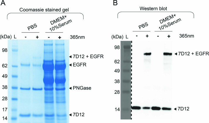
Extended Data Fig. 10. Photocrosslinking of 7D12-32pcY-109Bpa to EGFR performed in DMEM media containing 10% serum.
(A) The Coomassie stained gel demonstrates successful photocrosslinking of 7D12-32pcY-109Bpa to EGFR in phosphate buffered saline (PBS). For the same reaction performed in serum containing media, bands corresponding to sEGFR and photocrosslinked product are not clear on Coomassie stained gel due to the presence of other proteins in the serum. This is the full gel image for data shown in Fig. 5d. Lane marked L is the Invitrogen SeeBlue Plus2 Pre-stained Protein Standard (Catalog no. LC5925). These experiments were repeated twice with similar results. (B) The anti-His6 antibody western blot detects the C-terminal His6 tag on 7D12-32pcY-109Bpa. The photocrosslinked product, 7D12+ EGFR (top band in the blot) appears in lanes where 7D12-32pcY-109Bpa/ sEGFR in PBS or in serum containing media are irradiated with 365 nm light, demonstrating successful photocrosslinking under both conditions. The lower band corresponds to 7D12-32pcY-109Bpa/ 7D12-109Bpa and serves as a control demonstrating detection 7D12-32pcY-109Bpa/ 7D12-109Bpa in all samples. The image was acquired using GE ImageQuant™ LAS 4000 gel imager. This is the full gel image for data shown in Fig. 5d. Lane marked L is the Invitrogen SeeBlue Plus2 Pre-stained Protein Standard (Catalog no. LC5925). These experiments were repeated twice with similar results.

