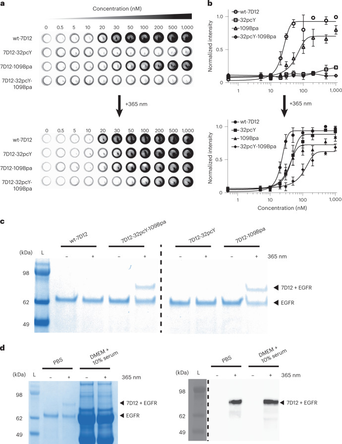
Fig. 5. Development of photoactive-photoreactive 7D12 mutant.
a, The on-cell binding assay demonstrates that 7D12-32pcY-109Bpa is a photoactive antibody. These experiments were performed in triplicate (Extended Data Fig. 8). b, The normalized intensities from the on-cell binding assay were plotted against the concentration of 7D12, where the x axis is in log10 scale. Each point in the graph represents mean values of normalized intensities ± s.d., designated as error bars, from three replicates. The data were fitted to the sigmoidal nonlinear equation using GraphPad to obtain binding affinity values (Kd). Before irradiation, the Kd values of wt-7D12 and 7D12-109Bpa were 23 (±2.6) nM and 54 (±14) nM, respectively. After irradiation, the Kd values of wt-7D12, 7D12-32Bpa, 7D12-109Bpa and 7D12-32pcY-109Bpa were 22 (±1.5) nM, 42 (±5.4) nM, 35 (±6) nM and 103 (±25) nM, respectively (Extended Data Fig. 8). For 7D12-32Bpa and 7D12-32pcY-109Bpa before irradiation, lines show connection between individual points. For all other experiments, lines show the fitting trace. c, Photocrosslinked product observed only with 7D12-109Bpa and 7D12-32pcY-109Bpa for samples irradiated with 365-nm light, demonstrating that 7D12-32pcY-109Bpa gets activated and then forms a covalent bond with EGFR upon irradiation with 365-nm light (Extended Data Fig. 9). These experiments were repeated twice with similar results. d, Photocrosslinking of 7D12-32pcY-109Bpa to sEGFR performed in DMEM containing 10% serum. The left panel shows Coomassie-stained gel demonstrating photocrosslinking of 7D12-32pcY-109Bpa to sEGFR in the control reaction performed in PBS. For the same reaction performed in serum-containing media, bands corresponding to sEGFR and photocrosslinked product are not clear on Coomassie-stained gel due to the presence of serum proteins (Extended Data Fig. 10). The right panel shows the anti-His6 western blot of the photocrosslinking reactions that detects the C-terminal His6 tag on 7D12 (Extended Data Fig. 10). The bands show the sEGFR–7D12 complex demonstrating successful photocrosslinking of 7D12-32pcY-109Bpa in serum-containing media. These experiments were repeated twice with similar results. For gel images, lanes marked L are the Invitrogen SeeBlue Plus2 Pre-stained Protein Standard (catalog no. LC5925).

