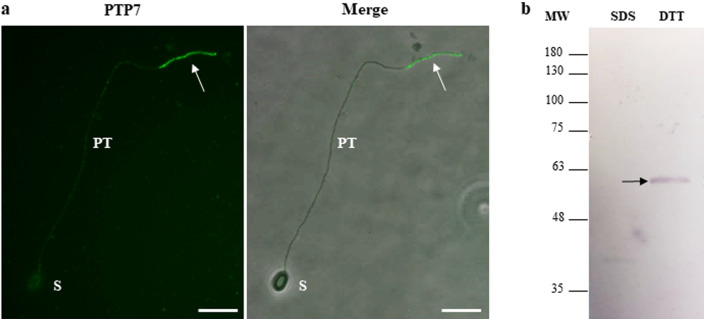
Figure 7.
Localization of the newly identified PTP7 at the terminal end of the extruded polar tube. (a) IFA with anti-PTP7 mouse antiserum (dilution 1:100) revealing a specific labelling of the terminal end of the A. algerae extruded polar tube (arrow). Secondary antibodies were Alexa 488-conjugated goat anti-mouse IgG. At least 10 different fields were observed which corresponds to ~ 50 to 130 spores with their polar tube extruded. See Supplementary Fig. S2 for additional pictures illustrating the different localizations of PTP7. PT polar tube; S spore; Scale bar: 10 µm. (b) Western Blot analysis on sporal proteins solubilized first in presence of 2.5% SDS (SDS extract) and then in a 100 mM DTT- containing solution (DTT extract). The antiserum reacted with a unique A. algerae sporal protein band at around 60 kDa in the DTT extract (arrow).

