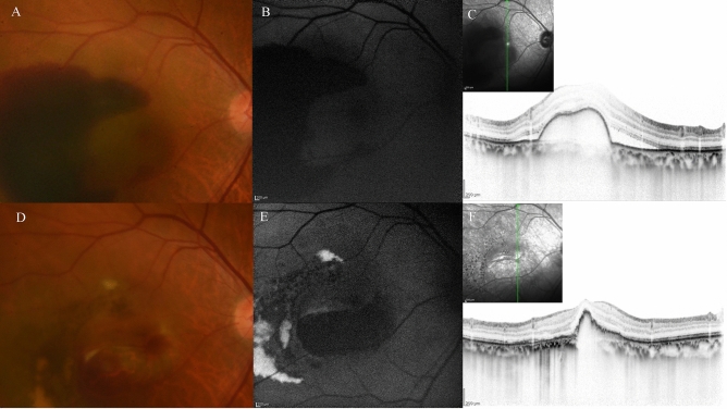
Figure 11.
A case of retinal pigment epithelial (RPE) tear in 77-year-old male patient with polypoidal choroidal vasculopathy. (A) Fundus photography revealed a haemorrhagic pigment epithelial detachment (PED) with subretinal fluid (SRF) and subretinal haemorrhage at the fovea. (B) Fundus autofluorescence (FAF) revealed no RPE tear at baseline (C) Optical coherence tomography (OCT) revealed the large haemorrhagic PED with SRF. (D) Fundus photography at 1 months after the third injection of faricimab revealed that RPE tear developed at the inferior of the PED. (E) FAF identified hemispherical RPE defect at the inferior part of the PED. (F) OCT revealed RPE defect at the inferior part of the pre-existing PED.SRF was completely absorbed. Regardless of the RPE tear development, visual acuity improved from 20/125 to 20/40.

