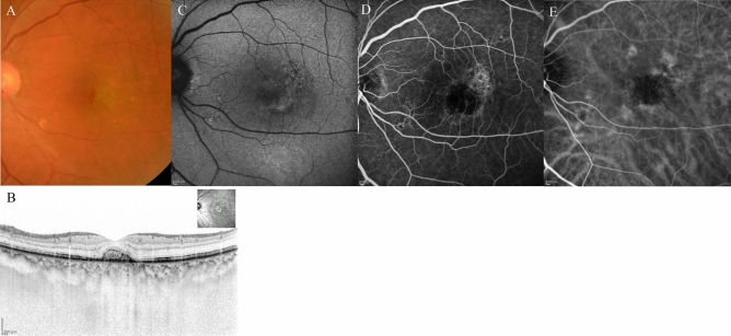
Figure 8.
The same 68-year-old male patient as shown in Fig. 7, here at 3 months after initiation of faricimab therapy. (A) Fundus photography shows absorption of the haemorrhage, fibrin, and yellowish mass. (B) Optical coherence tomography (OCT) revealed absorption of fibrin and haemorrhage, serous retinal detachment, and a small mass retained in the subretinal region. (C) Fundus autofluorescence imaging revealed very slight fine granular atrophy. (D) Late-phase fluorescein angiography detected no leakage from MNV. (E) Middle-phase indocyanine green angiography identified no abnormal vasculature.

