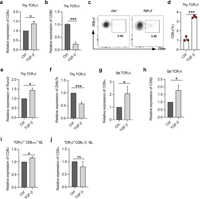
Fig. 5. TGF-β induces CD8α, but not CD8β expression in γδT cells.
a, b Relative expression of CD8α (a) and CD8β (b) to Hprt in overnight-cultured thymic γδT cells from C57BL/6 J mice in culture conditions of 1 μg/mL anti-CD3 combined with medium only (Ctrl), or in the presence of 2 ng/mL TGF-β1 (TGF-β) or 5 μM SB431542 (SB, TGF-β inhibitor), and detected by quantitative PCR. c Representative plot of 2-day-cultured thymic γδT cells from C57BL/6 J mice with CD8α staining in the presence of IL-2 (100 U/mL) based on culture condition of a to keep cells survive well in long-term culture. d Frequency of CD8α on thymic γδT cells from the same cells as in c. e, f Relative expression of Runx3 (e) and Zbtb7b (Th-Pok) (f) to Hprt in cells with the same culture condition as in a and detected by quantitative PCR. g, h Relative expression of CD8α (g) and CD8β (h) to Hprt on overnight-cultured splenic γδT cells from C57BL/6 J mice in the same culture condition as in a and detected by quantitative PCR. i, j Relative expression of CD8α to Hprt in overnight-cultured TCRγδ+CD8αα+ IELs (i) or TCRγδ+CD8α−β− IELs (j) from C57BL/6 J mice detected by quantitative PCR. *P < 0.05 and ***P < 0.001; ns no significant difference (unpaired two-tailed Student’s t-test). Data were representative of at least three independent experiments (means ± SEM).

