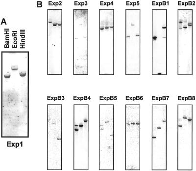
Figure 4.
Southern-blot analysis for Exp1-5 and ExpB1-8. A, Close-up view of the Southern blot for Exp1, indicating the pattern of loadings. B, Southern blots for Exp2–Exp5 and ExpB1–8 in the same pattern as shown in A. Ten micrograms of genomic DNA was digested with BamHI, EcoRI, or HindIII, separated on a 0.8% (w/v) agarose gel, and transferred to a nylon membrane. The blot was hybridized with a P32-labeled gene-specific probe using the same conditions as in Figure 3.

