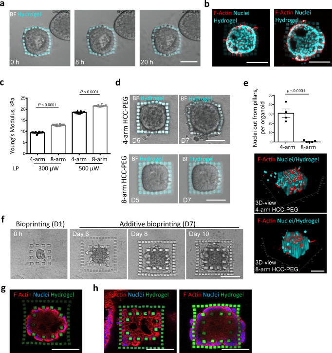
Fig. 3. Hydrogel-in-hydrogel live bioprinting for studying cancer cell migration in organoid 3D cultures.
a Brightfield time lap images (0, 8, 20 h) of a hydrogel-embedded tumor spheroid (day 6 post printing) growing within a cage of HCC-PEG pillars fabricated 1 day post organoid culture. Scale bar, 100 μm. b Fluorescent images of the tumor spheroid in c, showing different stages of cellular migration through the pillars day 7 post printing. Scale bars, 100 μm. c Young’s modulus measured by atomic force microscopy of 4-arm (black) or 8-arm (gray) HCC-PEG hydrogels photo-crosslinked at increasing laser power (300 or 500 μW) and wavelength (800 nm). Hydrogels were sequentially fabricated within the same Matrigel drop. Data are shown as mean ± s.d. of three independent replicates; all measurements performed are reported; unequal variance Student’s t-test was used; P < 0.05 was considered statistically significant. d Brightfield time lapse images of hydrogel-embedded tumor spheroid 5 or 7 days after bioprinting of 4-arm (upper panels) or 8-arm (lower panels) HCC-PEG pillars fabricated 1 day of organoid culture. Scale bars, 100 μm. e Quantification of nuclei detected out of the pillars of the hydrogel-embedded tumor spheroid 7 days after bioprinting of 4-arm (black) or 8-arm (gray) HCC-PEG pillars fabricated 1 day of organoid culture. Representative images of 3D reconstructions with xyz coordinates and 50 μm scale bar are shown. f Four-arm HCC-PEG hydrogel pillars were fabricated 1 day or 7 days post organoid culture (days 0 and 6 post printing, respectively) around a hydrogel-embedded growing tumor organoid. The brightfield images show the growth of the caged tumor spheroid at different time points. Scale bar, 100 μm. g Representative fluorescent image of tumor spheroid as in e at 14 days from first bioprinting step (day 15 of organoid culture). Scale bar, 100 μm. h Representative fluorescent images of cancer cells protruding through the bars of the first bioprinted hydrogel cage, invading the surrounding space and migrating through the pillars of the second fabrication step. Scale bars, 100 μm.

