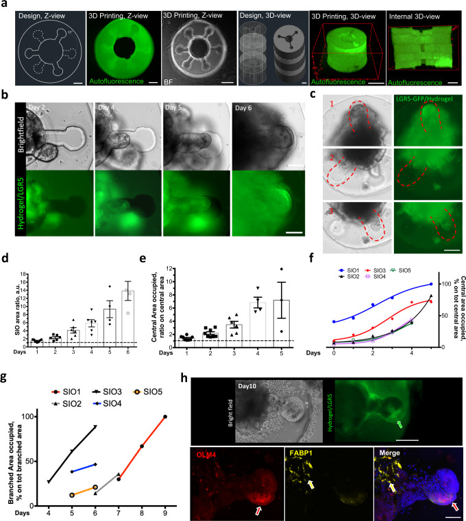
Fig. 4. Supra-organoid driven intestinal organoid morphogenesis via hydrogel-in-hydrogel live bioprinting.
a Representative bright field and fluorescence images showing 60°primordial small intestine design and HCC-gel hydrogels (left panel, top view; right panel, 3D reconstruction view). Scale bars 200 μm. b Representative bright field (upper) and fluorescence (lower) images of mSIOs just after primordial small intestine HCC-gel hydrogel printing or 2, 4, 5 or 6 days of culture and bioprinting. Budding was observed according to the defied shape of the hydrogel. Scale bars 100 μm. c Representative bright field and fluorescence images showing mSIO buds invading multiple crypts at different Z-levels of the primordial small intestine design after 6 days of culture post-printing. Scale bar 200 μm. d Quantification of the ratio between the area of the organoid at day 0 of culture (dashed line) and the area of the organoid during the following culture days (1–6 days). Statistical analysis is shown in Supplementary Table 1. e Ratio between the central area of the primordial intestine-shaped hydrogels with the organoid at seeding time (dashed line) and the area occupied by the organoid from day 1–6 of culture. Statistical analysis is shown in Supplementary Table 2.f Quantification of the percentage of central area occupied by the mSIOs during the culture (0–6 days). Calculation of the percentage was shown for 5 independent mSIO cultures. g Quantification of the percentage of branched areas occupied by 5 independent mSIO during the cultures (4–9 days). h Upper panels, representative bright field (upper) and fluorescence (lower) images showing mSIO budding after 10 days of culture within the primordial small intestine-shaped HCC-gel hydrogel. The arrow points at the LGR5 (green) cells. Lower panels, representative images showing immunofluorescence analysis for OLM4 (red) (corresponding to the LGR5-GFP in (i) and FABP1 (yellow) of mSIO cultured for 10 days within the primordial small intestine-shaped HCC-gel hydrogel). Nuclei are stained with Hoechst (blue). The arrows point at the branched (red) or central (yellow) portion of the mSIO in respect to the hydrogel. Scale bars 100 μm (upper panels), 50 μm (lowe panels).

