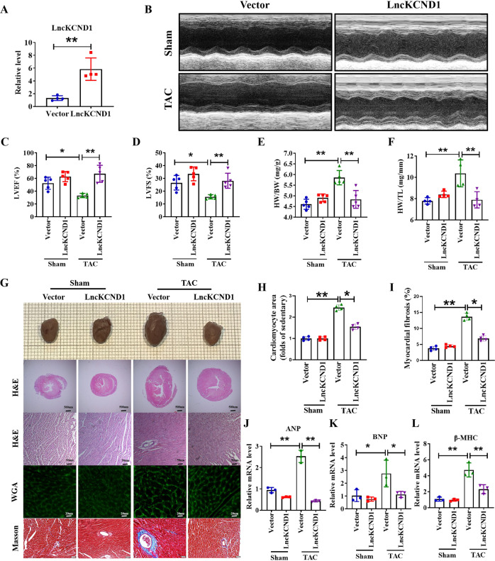
Fig. 3. Overexpression of LncKCND1 mitigates pressure overload-induced cardiac hypertrophy in vivo.
A LncKCND1 overexpression mice were generated by tail vein injection of adeno-associated virus serotype 9 (AAV9) carrying a vector or LncKCND1 for 4 weeks, and then subjected to Sham or TAC operation for 4 weeks (n = 4, **P < 0.01 vs. Vector). B–D Cardiac function of mice were determined by two-dimensional M-mode echocardiography (ECG). ECG assessment of left ventricle ejection fraction and fractional shortening of Sham or TAC mice infected with AAV9-Vector or AAV9-LncKCND1 (n = 5, *P < 0.05, **P < 0.01). E–F Comparisons of heart weight (HW)/body weight (BW) and HW/tibia length (TL) of Sham or TAC mice infected with AAV9-Vector or AAV9-LncKCND1 (n = 4-5, **P < 0.01). G Histological analysis of heart sections from Sham-Vector, Sham-LncKCND1, TAC-Vector and TAC-LncKCND1 groups. Five-µm-thick heart sections were stained with hematoxylin-eosin (H&E), wheat germ agglutinin (WGA) and Masson. H, I Statistics of cardiomyocyte size and myocardial fibrosis area (n = 4, *p < 0.05, **p < 0.01). J–L Relative mRNA levels of ANP, BNP and β-MHC in the heart tissues of Sham-Vector, Sham- LncKCND1, TAC-Vector and TAC- LncKCND1 groups (n = 3, *p < 0.05, **p < 0.01).

