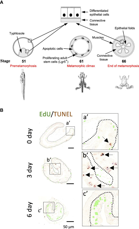
Figure 1.

(A). Schematic diagram of Xenopus intestinal metamorphosis. Both tadpole and frog intestine are structurally simple, consisting of mainly three tissue layers: inner epithelium, connective tissue, and outer muscle layers. The tadpole intestine is much simpler, with only a single epithelial fold, the typhlosole. In contrast, the frog intestine has multiple epithelial folds with elaborate connective tissue and muscle layers. The major events underlying the change from tadpole to frog intestine during metamorphosis include the apoptosis of essentially all larval epithelial cells, as indicated by circles. Concurrently, the adult epithelial stem cells, with high level expression of known stem cell markers such as Lgr5, are formed de novo through dedifferentiation of some larval epithelial cells and rapidly proliferate at the climax metamorphosis, as indicated by the dots. The connective tissue and muscle cells also develop extensively during metamorphosis. (B). Apoptotic and proliferating cells are non-overlapping epithelial cells during T3-induced intestinal metamorphosis. Premetamorphic Xenopus laevis tadpoles at stage 54 were treated with 10 nM T3 for 0, 3, or 6 days and sacrificed one hour after injection with EdU to label proliferating cells. Intestinal cross-sections were double stained for EdU and by TUNEL for apoptotic cells. A higher magnification of the boxed areas labeled with a’-c’ is shown on the right. The dotted lines depict the epithelium-mesenchyme boundary. Note that apoptosis as shown by the TUNEL signal in the epithelium occurred prior to the appearance of the clusters (islets) of EdU-labeled cells and in distinct epithelial cells during T3 treatment (c’). Arrows point to some apoptotic cells. See (11) for more details.
