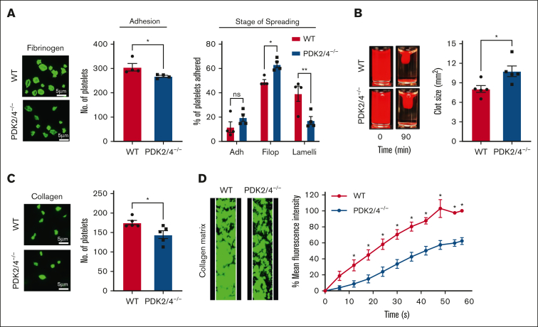
Figure 2.
PDK2/4−/− platelets exhibit reduced outside-in signaling and in vitro thrombus formation. (A) WT or PDK2/4−/− washed platelets (2 × 10⁷ cells/mL) stained using Alexa-Fluor 488-conjugated phalloidin were stimulated with PAR4 peptide (70 μM) for 10 minutes and added onto fibrinogen-coated (100 μg/mL) coverslips for 120 minutes. Five images were captured of each sample at random locations. A representative image of platelet adhesion and spreading is shown. Spreading platelets were divided into 3 classes: adhered (Adh) but not spread, filopodia (Filop): spreading platelets and lamellipodia (Lamelli): fully spread. Results are expressed as a percentage of the total number of platelets adhered. Cumulative data of the number of platelets adhered in each sample is shown. Values are mean ± SEM, n = 4 mice per group. Statistical analysis: Mann-Whitney U test (adhesion) or 2-way ANOVA (stage of spreading) followed by Sidak multiple comparisons test; ∗P < .05 and ∗∗P < .01. (B) Clot retraction was measured for 90 minutes in PRP, supplemented with red blood cells, after adding 1 U/mL of thrombin. The left panel shows the representative images at 90 minutes, and the right panel shows the quantification of the clot size. Values are mean ± SEM, n = 5 mice per group. Statistical analysis: Mann-Whitney U test; ∗P < .05. (C) WT or PDK2−/− washed platelets (2 × 10⁷ cells/mL) stained using Alexa-Fluor 488-conjugated phalloidin were added onto collagen-coated (100 μg/mL) coverslips for 45 minutes. Five images were captured of each sample at random locations. A representative image of platelet adhesion is shown. Cumulative data of the number of platelets adhered in each sample is shown. Values are mean ± SEM, n = 5 mice per group. Statistical analysis: Mann-Whitney U test; ∗P < .05. (D) DiOC6-loaded whole blood from WT or PDK2/4−/− mouse was perfused over a collagen-coated (100 μg/mL) matrix for 60 seconds at an arterial shear rate in a BioFlux Microfluidic flow chamber system. The left panel shows the representative image at the end of the assay, and the right panel shows the thrombus growth on the collagen matrix over time. Values are mean ± SEM, n = 3 mice per group. Statistical analysis: 2-way ANOVA followed by Sidak multiple comparisons test; ∗P < .05.

