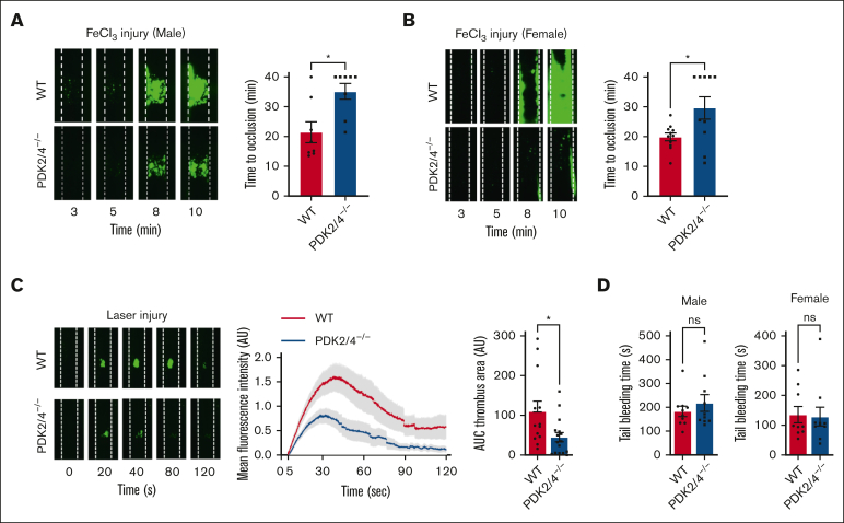
Figure 4.
PDK2/4−/− mice are less susceptible to experimental arterial thrombosis. (A) A representative image of carotid artery thrombus (5% FeCl3 injury for 2 minutes) and the time to occlusion in WT and PDK2/4−/− male mice is shown. The time to occlusion was measured until 40 minutes, the cutoff point at which the experiment was terminated. Values are mean ± SEM, n = 8 vessels from 8 mice per group. Statistical analysis: Mann-Whitney U test; ∗P < .05. (B) A representative image of carotid artery thrombus (5% FeCl3 injury for 2 minutes) and the time to occlusion in WT and PDK2/4−/− female mice is shown. Values are mean ± SEM, n = 10 to 11 vessels from 6 mice per group. Statistical analysis: Mann-Whitney U test; ∗P < .05. (C) A representative image of laser injury–induced mesenteric artery thrombus in WT and PDK2/4−/− male mice as visualized by upright intravital microscopy. The mean fluorescence intensity over time and AUC (thrombus area) is shown. Values are mean ± SEM, n = 13 to 16 thrombi from 4 mice per group. Statistical analysis: Mann-Whitney U test; ∗P < .05. (D) The tail-bleeding time in WT and PDK2/4−/− male and female mice was determined by measuring the time taken for the initial cessation of bleeding after the tail transection. Values are mean ± SEM, n = 10 mice per group. Statistical analysis: Mann-Whitney U test.

