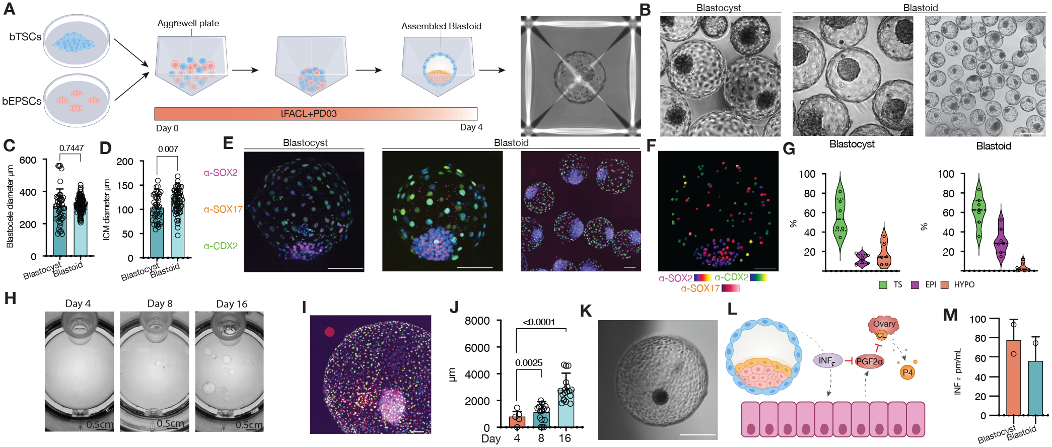
Figure 1. Assembly of bovine blastoids from EPSCs and TSCs cultures.

(A) Illustration of the assembly process via bovine EPSCs and TSCs aggregation. (B) Phase-contrast image comparing blastoids vs blastocysts. (C) Blastocele diameter measurement. (D) Inner cell mass (ICM) diameter measurement. (E) Immunostaining for epiblast marker SOX2 (magenta, EPI), hypoblast marker SOX17(red, HYPO) and trophectoderm marker CDX2(green, TS), individual markers in Figure S1. (F) Blastocyst and Blastoid lineage composition quantified via confocal microscopy 3D reconstruction and spots colocalization quantification using IMARIS. (G) Snapshots of in vitro growth of blastoids in a rotating culture system (Clinostar Incubator, Celvivo). (H) Representative image via immunostaining of all three lineages as in e, individual markers in Figure S4. (I) Blastoid diameter quantification. (J) representative micrographs of in vitro grown blastoid. (K) A schematic of the maternal recognition of the action of pregnancy signal interferon TAU (INFt). (L) Enzyme-linked immunosorbent assay (ELISA) measurement of (INFt) in surrogate recipients following embryo transfers. PGF2α: Prostaglandin F2α. CL: Corpus luteum. P4: Progesterone.
