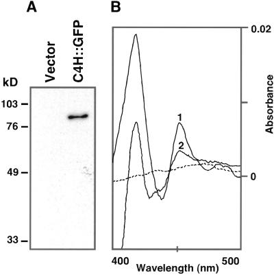
Figure 5.
Immunoblot and carbon monoxide-induced differential absorption spectra analysis of microsomes from C4H::GFP-transformed yeast. A, Immunoblot analysis of control (vector only) and experimental (C4H::GFP-transformed) yeast strains. Microsomal protein preparations (2 μg) were separated by SDS-PAGE and transferred to a PVDF membrane. The blot was incubated with mouse monoclonal anti-GFP antibody, and signals detected by chemiluminescence. B, Carbon monoxide-induced differential absorption spectra recorded from reduced microsomes of C4H-GFP transformed yeast. Microsomes from 20 or 12 h Gal-induced C4H::GFP- transformed yeast, adjusted to a final assay concentration of 1.0 or 1.1 mg/mL, were used to record spectrum 1 or 2, respectively. The spectrum for microsomes from vector transformed yeast is shown by the dashed line.

