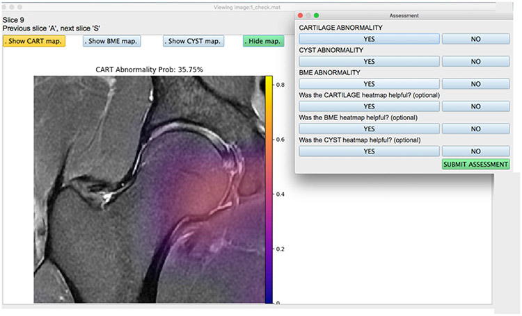
FIGURE 3:
View of the user-friendly AI-based assist tool for radiologists. Images were completely stripped of their metadata and any patient information and loaded onto the tool. The radiologist has the option of turning on or off the cartilage lesions, bone marrow edema-like lesions, and subchondral cyst-like lesion saliency maps generated on the test set using the model. Once the assessment was submitted, the responses were recorded and stored for each patient image. The radiologist can select from a list of patient images, where each file is simply labeled with a number, and corresponding grades are mapped onto the Excel sheet.

