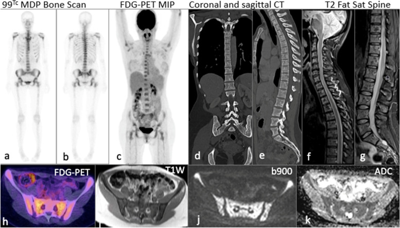
Fig. 3.
45-year-old female with multifocal grade 2 invasive ER positive HER2 negative metastatic lobular breast cancer: 99m Tc MDP planar bone scan (a, b) and coronal maximum intensity projection PET/CT (c) show no uptake in the axial or appendicular skeleton. Coronal and sagittal CT reformats (d, e) performed 1 week later demonstrating subtle sclerotic changes. Sagittal fat saturated T2W sequence of the spine (f, g) shows heterogenous marrow signal. Fused axial PET/CT (h) shows no FDG avidity. Widespread bone metastases in the same patient on WB DW-MRI (i) axial T1W (j) b900 and (k) ADC

