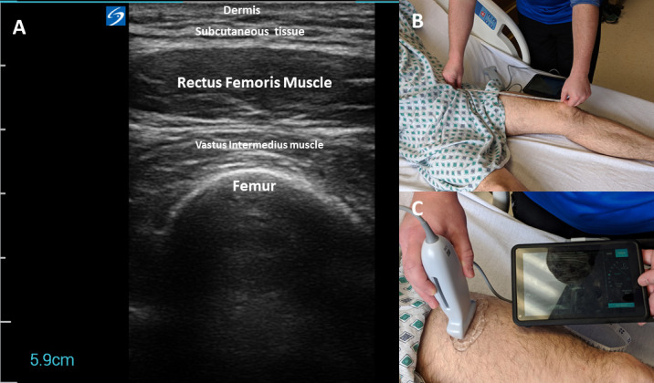
Figure 1.

Representative images of ultrasound acquisition techniques and the obtained image of the rectus femoris muscle. (A) is a representative ultrasound image with anatomical structures labelled for the quadriceps muscle. (B) demonstrates the technique to locate the anatomical landmarks for rectus femoris ultrasound (two-thirds of the distance from anterior superior iliac spine to the superior border of the patella). (C) depicts the-minimal-to-no-compression technique using the ultrasound probe with adequate ultrasound transmission gel to obtain images. These images were staged by the authors to demonstrate appropriate technique and were not taken from a patient encounter.
