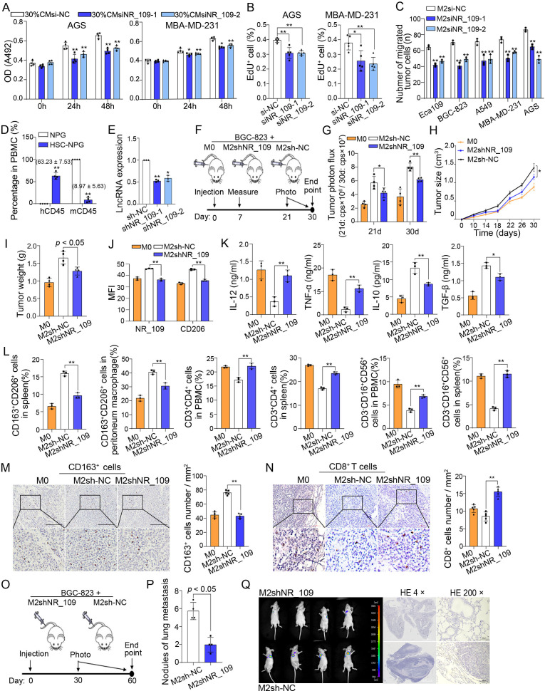
Figure 2.
Knockdown of NR_109 reduced the activity of M2-like macrophages to promote growth and metastasis of cancer cells. (A) The MTS and (B) EdU incorporation assays showed the proliferation of tumor cells in the coculture system with 30% culture medium (CM) of M2-NR_109low. (C) The migration of tumor cells when cocultured with M2-NR_109low cells. (D) The proportion of hCD45+ cells in the PBMC of NPG mice and HSC-NPG mice (8.97% ± 5.63% vs. 63.23% ± 7.53%). (E) Expression of NR_109 in M2-like macrophages after NR_109 lentiviral transduction was measured by qPCR. (F) The sketch of subcutaneous xenograft model in HSC-NPG mice. (G) The tumor growth (shown as photon flux) was examined by in vivo imaging. (H) The tumor size, (I) tumor weight and (J). MFI of NR_109 and CD206 in tumor tissues of different groups were analyzed. (K) The level of IL-12, TNF-α, IL-10 and TGF-β in the serum of HSC-NPG mice from distinct groups was tested by ELISA. (L) The percentage of M2-like macrophages (CD163+CD206+) in spleen and peritoneum macrophages, and the percentage of CD4+ T cells and NK cells (CD3-CD16+CD56+) in the PBMC and spleen were measured by FCM. (M) The infiltration of CD163+ cells and N. the number of CD8+ T cells in tumor tissues of different groups was detected by using IHC assays.(O) The sketch of metastatic tumor model in nude BALB/c mice. (P) The number of lung metastasis nodules was examined. (Q) In vivo imaging and HE staining showed the lung metastasis nodules and the representative regions of the lung in HSC-NPG mice of the two groups. The statistical data are from three independent experiments and the bar indicates the SD values (*p < 0.05, **p < 0.01). MFI, mean fluorescence intensity.

