FIG 2.
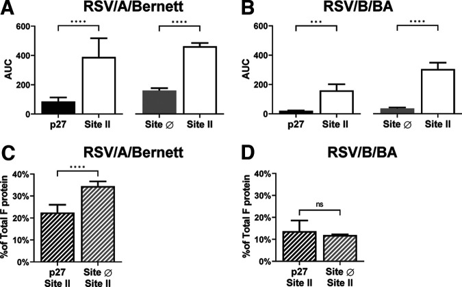
p27 and F protein in the prefusion conformation can be detected at quantifiable amounts on the surface of spRSVs. Areas under the curve (AUC) of paired measures of p27 and site II or site Ø and site II probed with monoclonal antibody palivizumab (anti-site II), RSV7.10 (anti-p27), or D25 (anti-site Ø) for spRSV/A/Bernett (A) and spRSV/B/BA (B) are shown. Site II was used as a surrogate for total F protein, and site Ø is a pre-F-specific antigenic site. Ratios of the AUC of p27 to site II and site Ø to site II determined relative proportions of p27 and pre-F to total F protein on spRSV/A/Bernett (C) and spRSV/B/BA (D). Error bars are standard deviations (n = 4 replicates). Statistical significances were determined by unpaired parametric t test in GraphPad Prism, assuming Gaussian population distribution. Correlations were calculated with a two-tailed test. P values of <0.05 were considered significant, with a 95% confidence interval. ****, P < 0.0001. ns, not significant.

