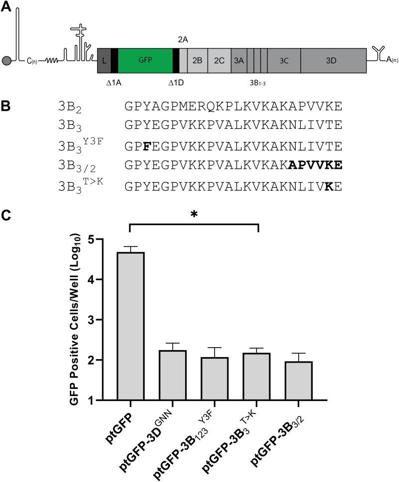
FIG 1.
A single amino acid substitution at the 3B3-3C junction prevents FMDV replicon replication. (A) Schematic diagram of the FMDV replicon. (B) Sequence alignments of the 3B cleavage junctions with the 3B1,2,3Y3F, 3B3/2, and 3B3T>K mutants. (C) Replication of replicons containing 3B3 mutations as well as the WT ptGFP replicon (ptGFP) and replication-defective controls containing inactivating mutations in 3Dpol (3DGNN), or 3B proteins (3B1,2,3Y3F). GFP expression was monitored hourly for 24 h. The graph shows GFP positive cells per well at 8 h posttransfection when replication is maximal. Significance compared to WT control (n = 3 ± SEM; * = P ≤ 0.05).

