Figure 7.
Continued.
Figure 7.
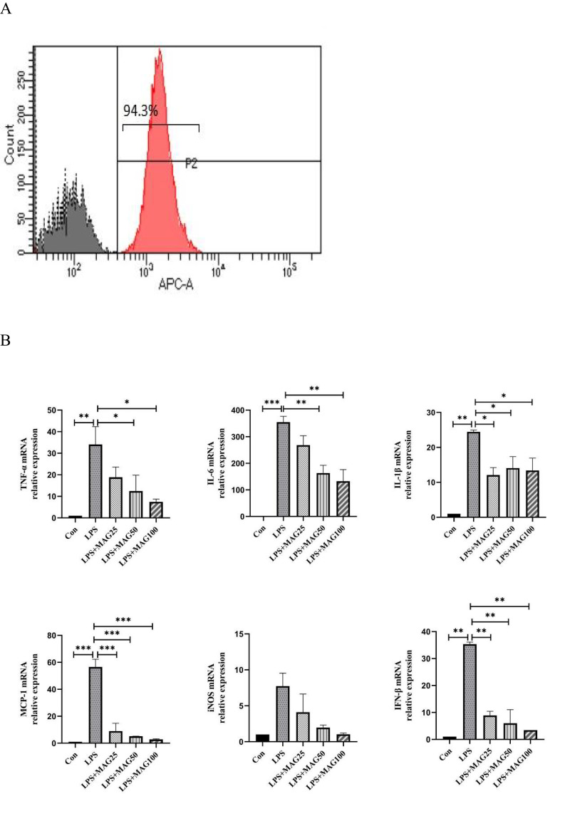
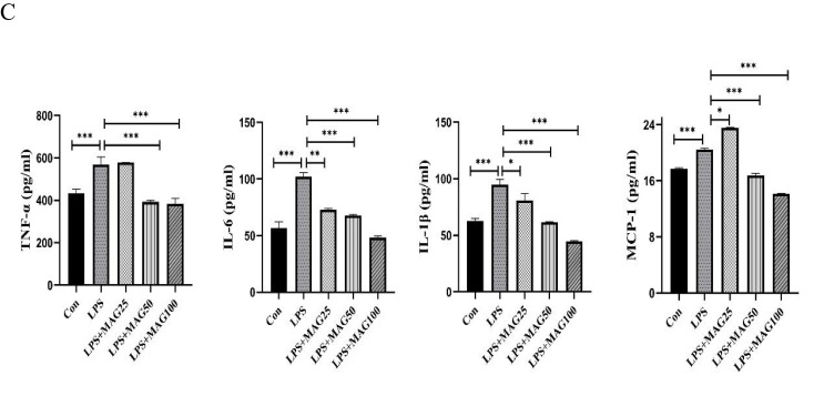
The effects of Mag on the pro-inflammatory cytokines, mediators, and chemokines of LPS-stimulated peritoneal macrophages. (A) The proportion of macrophages in the peritoneal cells from mice were detected by flow cytometry. Representative overlay histograms. Gray-filled histograms indicate isotype control. (B) The mRNA expression of TNF-α, IL-6, IL-1β, MCP-1, iNOS, and IFN-β mRNA in LPS-induced peritoneal macrophages were determined by qRT-PCR. (C) ELISA was used to detect the concentrations of TNF-α, IL-6, IL-1β, and MCP-1 in the peritoneal macrophages. Data in (A–C) are representative of at least three repetitions. *P<0.05, **P<0.01, ***P<0.001 (one-way ANOVA with Tukey’s post hoc test or Kruskal–Wallis test).
Abbreviations: Mag, magnoflorine; ELISA, enzyme linked immunosorbent assay; TNF-α, tumor necrosis factor; IL-6, interleukin-6; IL-1β, interleukin-1β; MCP-1, monocyte chemoattractant protein-1; iNOS, inducible nitric oxide synthase; IFN-β, interferon-beta; qRT-PCR, Quantitative Real-Time PCR; LPS, lipopolysaccharide.


