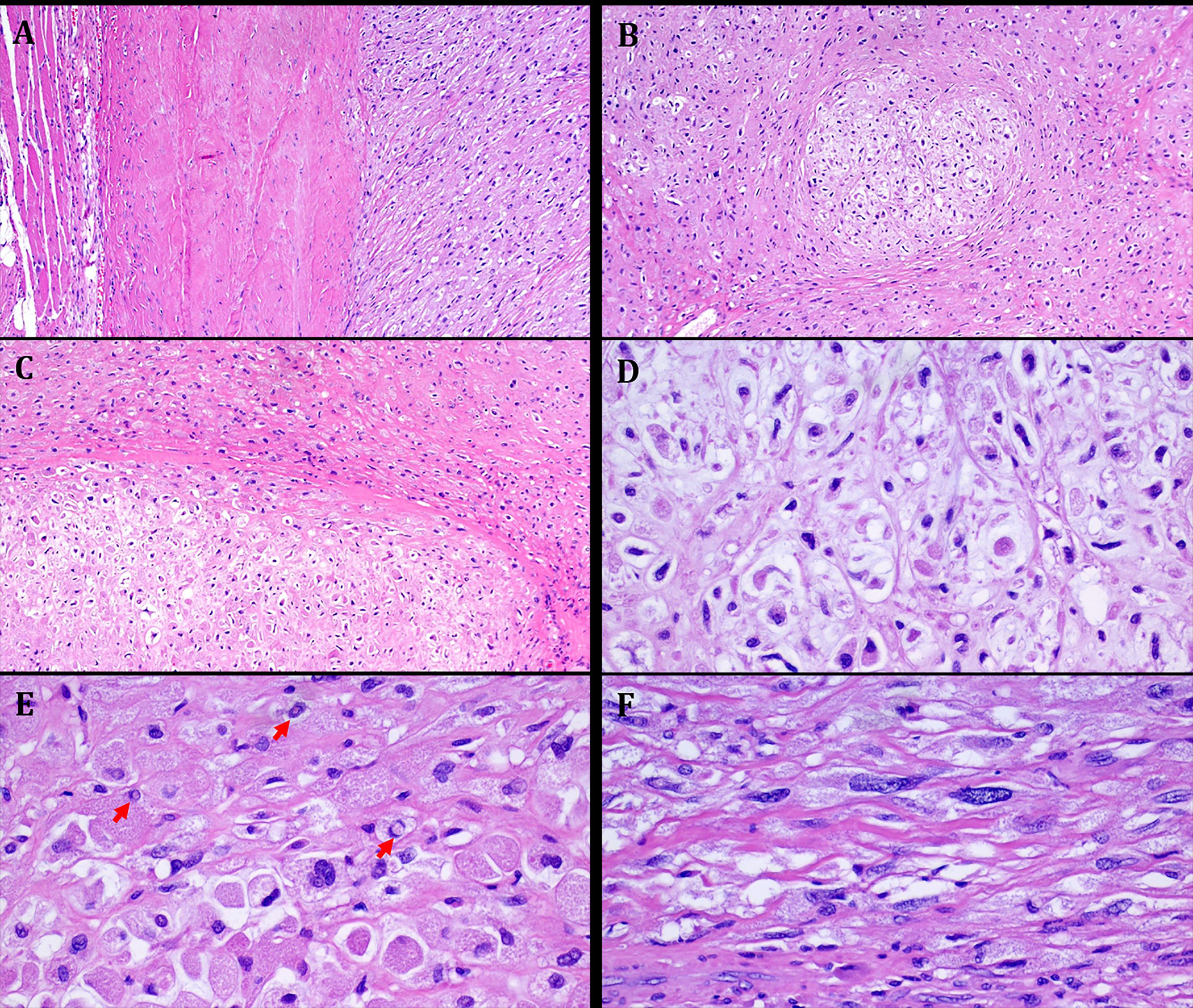
FIGURE 3.

Representative images illustrating the histological features in Case 2. A. well-circumscribed tumor periphery surrounded by fascia and muscle on left. B: at low power, chondromyxoid lobules are seen embedded within hyaline variably sclerosed stroma. C: higher magnification showing abrupt transition between chondromyxoid and hyaline areas. D: admixture of large epithelioid variably vacuolated tumor cells and scattered spindle cells seen at high power. E: prominent eosinophilic and granular cytoplasmic quality, note numerous nuclear pseudoinclusions (arrows). F: scattered spindle cells with hyperchromatic atypical looking nuclei.
