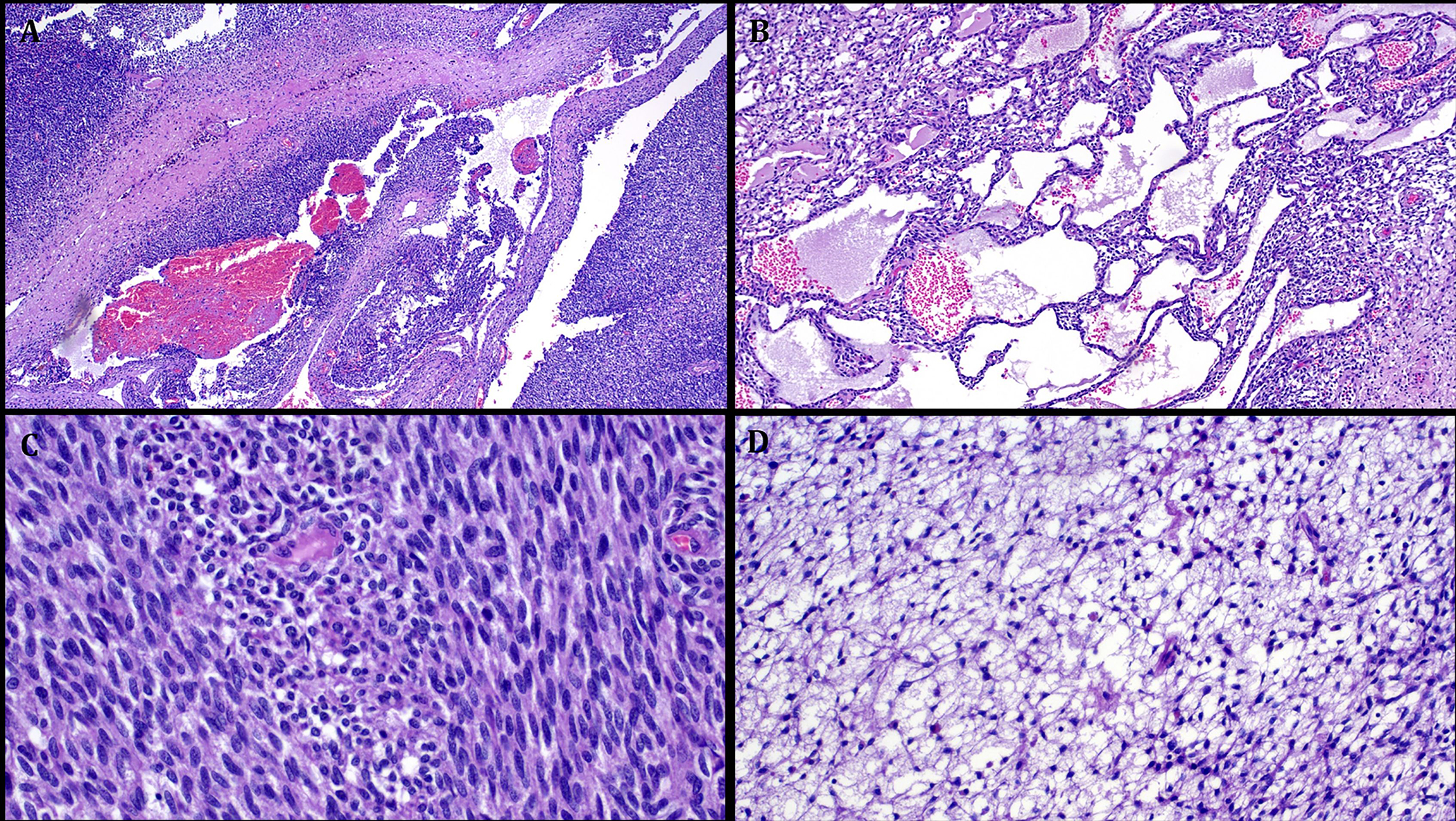
FIGURE 5.

Representative images illustrating the histological features in Case 4. A: low power view showing highly cellular neoplasm with alternating solid and cystic areas. B: prominent macrocystic stromal degeneration. C: highly cellular fibrosarcoma-like areas (with little to no mitoses). D: prominent primitive reticular-myxoid stromal features reminiscent of EMCMT of tongue.
