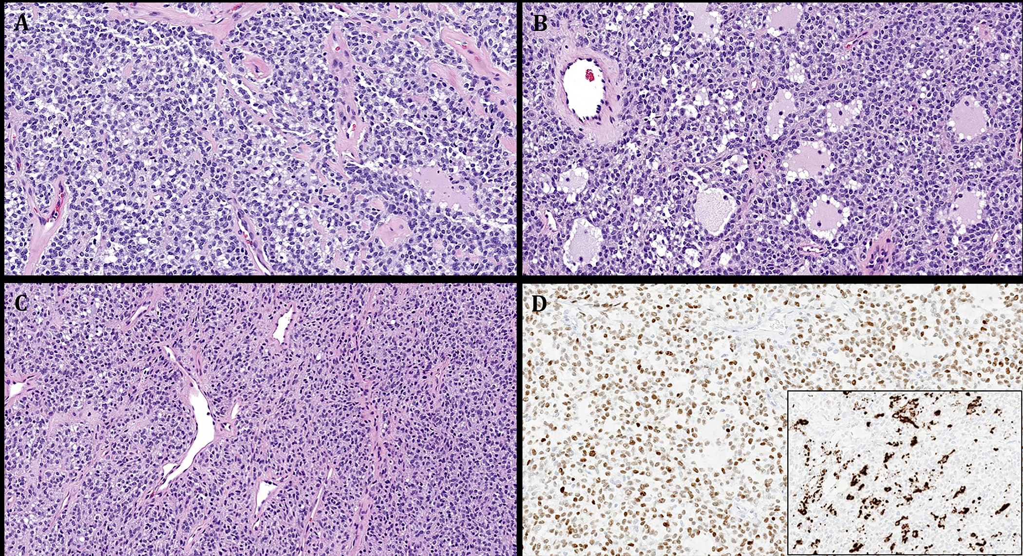
FIGURE 6.

Representative images illustrating the histological features in Case 5. A: monotonous round to ovoid bland cells arranged into non-descript confluent sheets. B: prominent pseudofollicular spaces are seen. C: cellular spindle cell areas with thin-walled gaping vessels. D: main image: diffuse expression of MyoD1. Subimage: variable expression of desmin.
