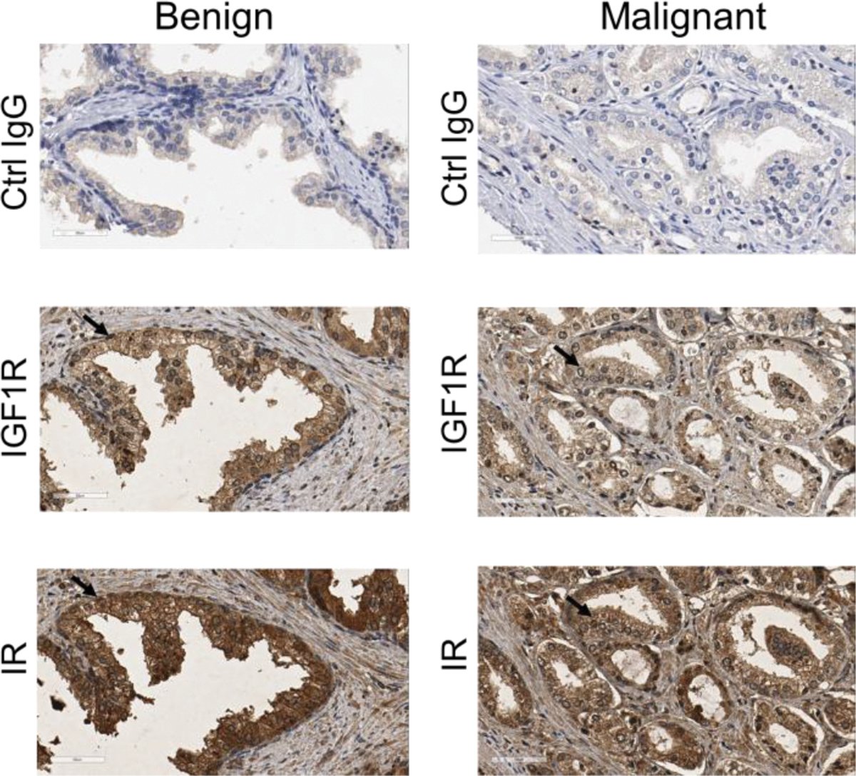
Fig. 9.

Expression of IGF1R and IR in prostate benign and malignant tissue using immunohistochemistry (IHC). Control IgG, a non-immuned mouse IgG was diluted to match the IgG concentration of IGF1R and IR antibodies for negative control. Arrows indicated positive cells.
