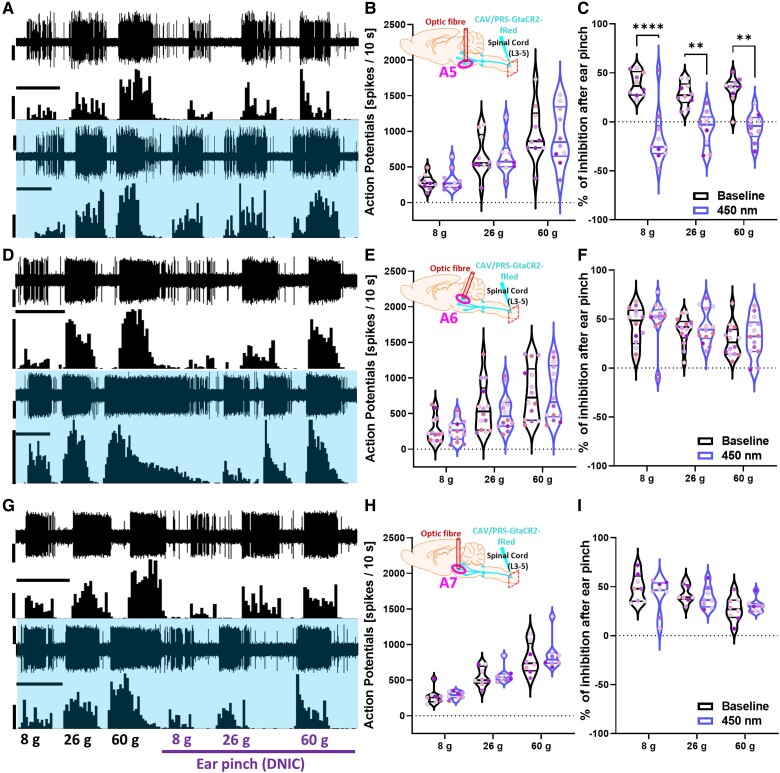
Figure 3.
Inhibition of spinally projecting A5 neurons abolishes DNIC. (A, D and G) Example traces of DDH-WDR evoked neuronal firing before and after GtACR2-mediated inhibition (450 nm continuous laser light illumination, 400 mW/mm2) of labelled A5, A6, and A7 nuclei, respectively. (B, E and H) The basal evoked response of DDH-WDR neurons were not altered upon optical inhibition of A5, A6, or A7 nuclei. (C) DNIC were abolished after A5 GtACR2- but not A6 or A7 mediated inhibition, F and I, respectively. Data represents mean ± SEM. Dots represent individual neurons (A5: N = 6 rats, n = 10 neurons, A6: N = 7, n = 14, A7: N = 6, n = 8), and dots are colour coded to reflect neurons studied from the same animal. Two-way RM-ANOVA with Tukey post hoc: **P < 0.01, ****P < 0.0001. Scale bars in A, D and G: waveform trace = 60 μV; spike count = 60 spikes; time scale = 20 s. See Supplementary Figs 3 and 4.

