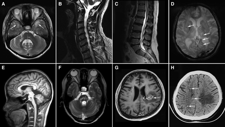
Figure 2.
Characteristic MRI features of genetic cerebral small vessel disease. (A) T2 axial image showed hyperintensities of the anterior temporal lobes (arrows) in a 47-year-old patient with CADASIL. (B) Cervical MRI showed spondylosis deformans in a 57-year-old male patient carrying a heterozygous HTRA1 mutation. His affected brother suffered long-term low back pain and lumbar MRI showed spondylosis deformans (C). (D) Susceptibility-weighted image showed intracranial haemorrhage in the left occipital cortex (arrow) and multiple cerebral microbleedings (CMBs, arrows) in a 66-year-old patient with COL4A2-related disorders. (E and F) T1 sagittal and T2 axial images showed fused infarctions (arrows) in the pons in a 43-year-old patient with pontine autosomal dominant microangiopathy and leukoencephalopathy (PADMAL). (G) Post-contrast MRI of a 41-year-old patient with retinal vasculopathy with cerebral leukodystrophy and systemic manifestations (RVCL-S) showed an enhanced lesion with a mass effect and surrounding oedema (arrow) in the left parietal white matter. Punctate calcifications (arrows) were detected in the patient by the CT scan (H).

