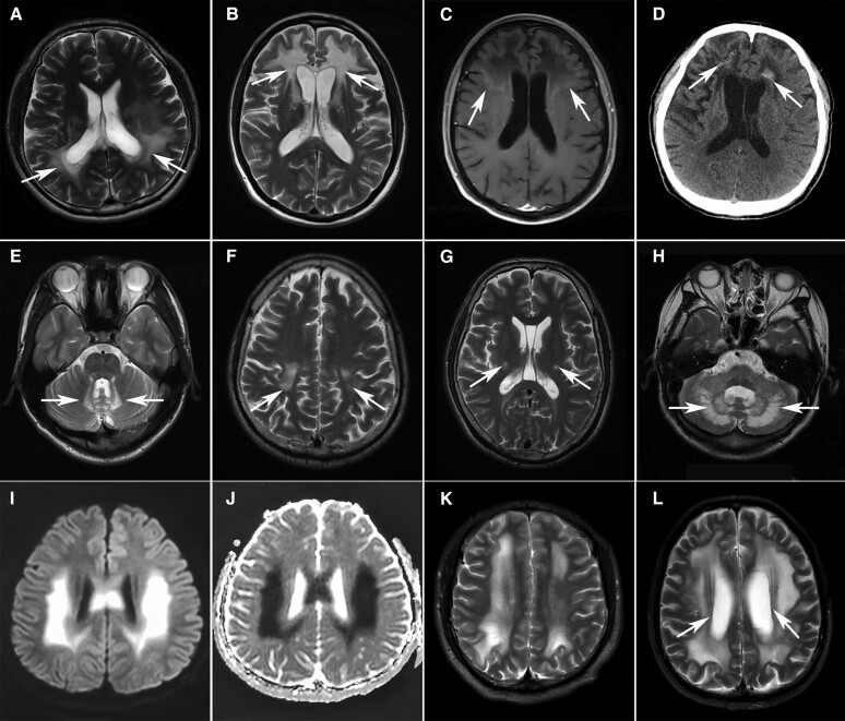
Figure 3.
Characteristic MRI features of myelin disorders. (A–E) Cerebral ALD with parieto-occipital (A, arrows) or frontal (B, arrows) predominance. Note the contrast enhancement (arrows) marginal to the demyelinated areas (C) and punctate calcification (arrows) in the lesions on CT scan (D). The spinocerebellar variant involved the bilateral cerebellar dentate nucleus (E, arrows). (F and G) Bilateral T2 hyperintensity of corticospinal tracts from the motor cortex (F, arrows) along the posterior limb of the internal capsule (G, arrows) to the brainstem in a 46-year-old patient with Krabbe disease. (H) T2-weighted hyperintensities in the bilateral cerebellar dentate nucleus (arrows) in a 36-year-old female patient with cerebrotendinous xanthomatosis (CTX). (I and J) Hyperintensities on diffusion-weighted imaging (I) and hypointensities on apparent diffusion coefficient imaging (J) in the bilateral parietal regions in a 21-year-old male patient with X-linked Charcot–Marie–Tooth disease (CMT-X). (K) Diffuse white matter lesions in parallel with the lateral ventricles in a 23-year-old patient with phenylketonuria (PKU). (L) Extensive T2-weighted hyperintensities in the subcortical and deep white matters with relative sparing of the periventricular rim (arrows) in a 66-year-old patient with adult-onset autosomal dominant leukodystrophy (ADLD).

