Figure 4.
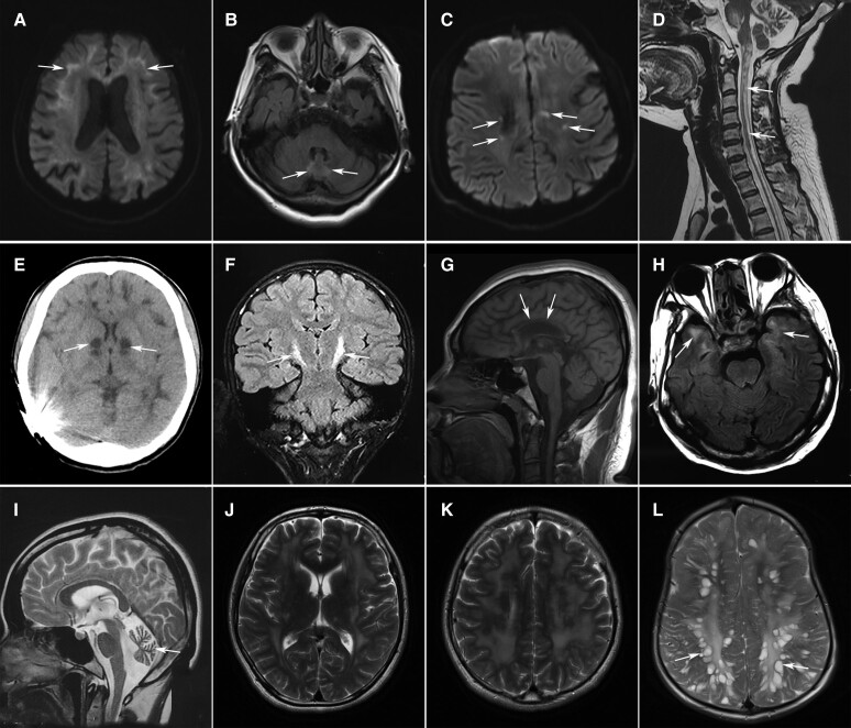
Characteristic imaging features of leuko-axonopathies. (A and B) High signals on diffusion-weighted imaging (DWI) along the corticomedullary junction (A, arrows) and T2-weighted hyperintensities in the cerebellar vermis (B, arrows) in a 65-year-old female with NIID. (C) Punctate hyperintensity (arrows) within the white matter lesions on DWI in a 29-year-old male with AARS2-related leukoencephalopathy. (D) Long-tract involvement of the spinal cord (arrows) in a 36-year-old female with leukoencephalopathy with brainstem and spinal cord involvement and elevated cerebrospinal fluid lactate (LBSL). Severe cerebellar atrophy was also noted. (E) Abnormal signals of the basal ganglia (arrows) and diffuse white matter lesions on CT scan in a 19-year-old male patient with mitochondrial disease. (F) Hyperintensities along bilateral corticospinal tracts (arrows) on coronal FLAIR images in a male patient with Pol-III-related disorder. (G) Severe atrophy of the corpus callosum (arrows) in a 25-year-old female with spastic paraplegia-15 (SPG15). (H) Diffuse white matter lesions with the involvement of temporal lobes (arrows) in a patient with myotonic dystrophy type 1. (I) Marked cerebellar atrophy in a 26-year-old female with Sandhoff disease. (J and K) Extensive white matter lesions in a 27-year-old male with Gordon Holmes syndrome. (L) Enlarged perivascular spaces resembling bunches of grapes in a male patient with mucopolysaccharidosis type II.

