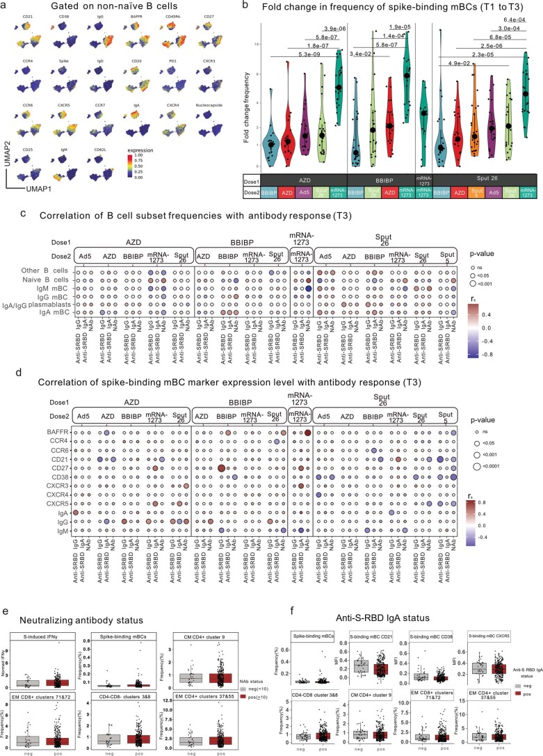
Extended Data Fig. 3. Analysis of the B cell compartment after vaccination.
a, UMAP showing the distribution of markers expressed by non-naïve B cells (IgD-/IgM-) among all vaccine groups (n = 799). b, Fold change of spike-binding mBC frequencies at T3 compared to the mean per group at T1 (n = 347). Large black dots show the median, and the vertical line spans the interquartile range. P-values were calculated using the Mann-Whitney-Wilcoxon test between groups with the same dose 1 and the Benjamini-Hochberg method to correct for multiple hypothesis testing. Only significant P values (P < 0.05) are displayed. c, Correlation among B cell subsets and antibody responses (anti-S-RBD IgG levels, anti-S-RBD IgA levels and neutralizing antibody titers) (n = 347). d, Correlation among spike-binding mBC marker expression levels and antibody responses. Color indicates the Spearman’s rank correlation coefficient (rs), and the circle size indicates the P value (n = 347). e, Participants were classified as negative (< 10) or positive (≥ 10) for neutralizing antibodies (n = 347). f, Participants were positive (>0.32 ng/ml) or negative for anti-S RBD IgA (n = 345). (e,f) The means of each listed parameter were compared between negative and positive responders using the Mann-Whitney-Wilcoxon test and corrected for multiple hypothesis testing with the Benjamini-Hochberg method. Boxes bound the interquartile range (IQR) divided by the median, and Tukey-style whiskers extend to a maximum of 1.5 × IQR beyond the box. Dots are participant data points.

