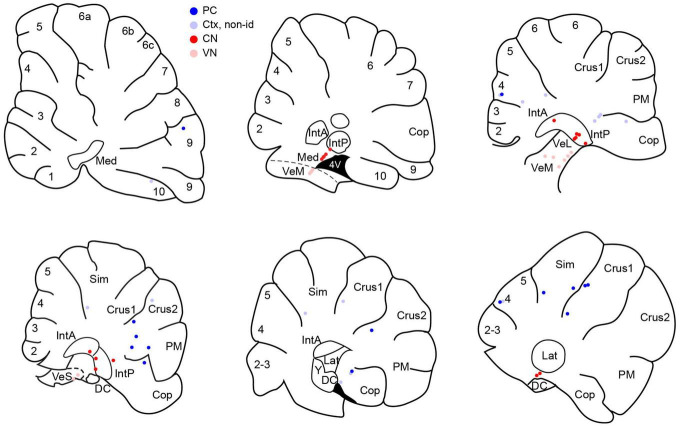
FIGURE 1.
Distribution of histologically-localized neurons. Schematics of parasagittal sections through the cerebellum at mediolateral distances going from approximately 0.5 to 3.5 mm from the midline. Dark blue circles indicate PCs. Pale blue circles are non-identified cortical neurons. Red circles indicate CN neurons and pink circles indicate VN neurons. Numbers on schematics correspond to lobule numbers. Cop, copula; DC, dorsal cochlear nucleus; IntA, IntP, anterior and posterior interpositus; Lat, lateral CN; Med, medial CN; PM, paramedian; Sim, simplex; VeL, VeM, VeS, lateral, medial, superior vestibular nucleus.

