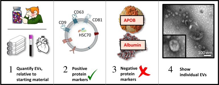
Figure 3.
Steps towards EV characterization, adapted from MISEV2018 guidelines.32 (i) Determine the quantity of EVs obtained, relative to the amount of starting material. (ii) Verify the presence of at least three positive protein markers of small EVs, including one transmembrane or GPI-anchored protein (e.g. CD9, CD63, CD81, NT5E/CD73), and one cytosolic, luminal protein (e.g. ALIX/PDCD6IP, HSC70). For large EVs, a wide range of surface markers such as integrins from the cell of origin may be used. (iii) Preferably, demonstrate the relative abundance of significant contamination by non-vesicular, co-isolated components such as lipoproteins (APOB, APOA1, APOA2) or albumin. (iv) Characterize individual EVs, with images of single EVs (both wide-field and close-up).

