
Fig. 7 |. Electrical interfaces with the central nervous system.
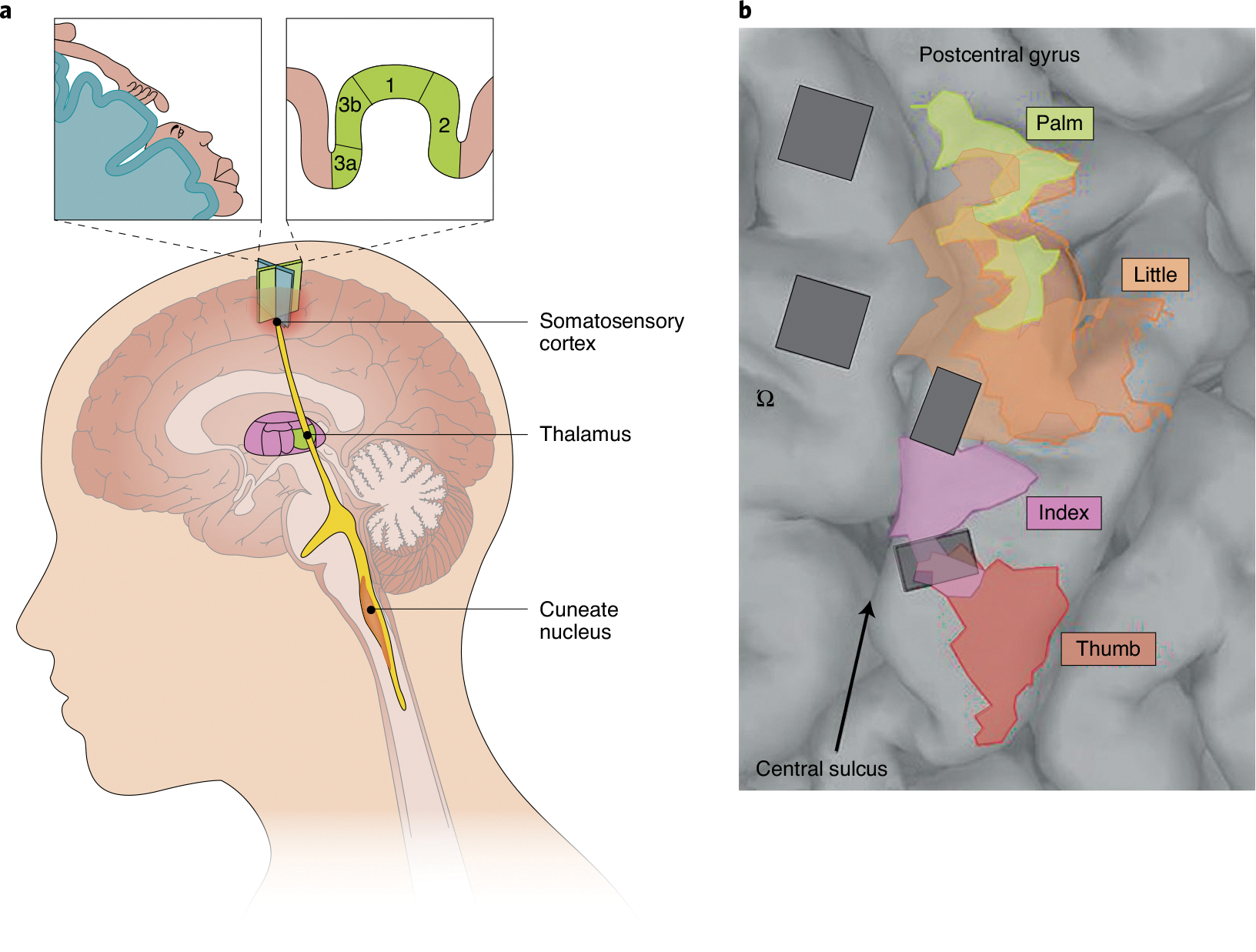
a, Possible loci of neural interfaces for the restoration of touch in the central nervous system. From bottom to top: the cuneate nucleus, the thalamus and the somatosensory cortex. The insets indicate the arm and face regions of the somatosensory homunculus in the human somatosensory cortex (top left), and the cortical fields in the Brodmann’s areas 3a, 3b, 1 and 2 of the somatosensory cortex (top right). b, Surgical image showing the implantation, in a human, of two arrays in the motor cortex (anterior to the central sulcus; left) and of two arrays in the somatosensory cortex (posterior to the central sulcus; right)146. The coloured areas indicate the mapping of the palm, thumb, index finger and little finger in the somatosensory cortex. The symbol ‘Ω’ indicates the presumptive location of the hand in the motor cortex (the so-called ‘hand knob’). Credit: Image courtesy of Kenzie Green; adapted with permission from ref. 146, AAAS