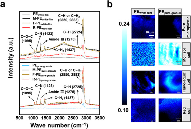
Figure 1.
Confocal micro-Raman spectroscopy analysis of the coating on microplastic particles incubated with the water extracted soil metabolites from a mollisol soil (M), a fluvo-aquic soil (F), and a red soil (R), respectively. (a) Raman spectra. (b) False color Raman image of the microplastic particles (dark) and the biomolecules forming a putative eco-corona (light) on their surfaces, generated from the spectral mapping data. The brightness of the color represents the relative percentage of eco-corona on the surface of microplastics; the lighter the color, the larger the percentage of eco-corona. Window size, 50 × 50 μm. PEwhite-film: white polyethylene film microplastics; PEpure-granule: pure polyethylene microplastic granules. The black polyethylene film microplastics cannot be analyzed by Raman detection because of the limitation of the detection technology.42

