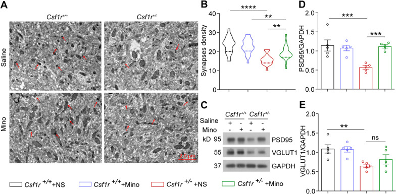
Fig. 7.
Minocycline partially restores synaptic density in forebrain tissue of Csf1r+/− mice. A Electron micrographs of synapse density in the CA1 area of forebrain sections from 9-month-old Csf1r+/+ or Csf1r+/− mice administered or not minocycline. B The number of synapses in the CA1 area of Csf1r+/+ or Csf1r+/− mice treated or not with minocycline was analyzed (n = 4 mice per group). C Western blot detection of postsynaptic membrane protein marker (PSD95) and presynaptic membrane protein marker (VGLUT1) in forebrain lysates of Csf1r+/+ or Csf1r+/− mice treated or not with minocycline. D PSD95 protein levels quantified by densitometry with GAPDH for comparison (n = 5 mice per group). E VGLUT1 protein levels quantified by densitometry with GAPDH for comparison (n = 5 mice per group). Data are presented as means ± SEM. P-values were calculated using two-way ANOVA post-Sidak’s multiple comparisons tests. **p < 0.01, ***p < 0.001, ****p < 0.0001, ns, no significance. Mino, minocycline

