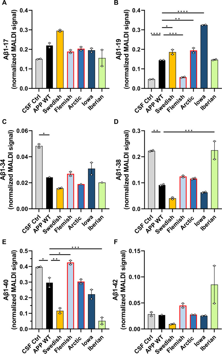
Fig. 11.
Comparison of secreted Aβ proteoforms from APP WT and APP FAD mutants. Normalized MALDI-TOF signals of A Aβ1-17, B Aβ1-19, C Aβ1-34, D Aβ1-38, E Aβ1-40 and F Aβ1-42 were plotted from the mass spectra of the APP WT and APP FAD mutant HEK293T cell media samples after IP-MALDI-TOF. The circles represent the two biological replicates (N = 2), each consisting of the average normalized value of two technical replicates from MALDI-TOF. Normalization was performed by dividing peak areas of each proteoforms by the sum of the peak areas of Aβ1-15, Aβ1-16, Aβ1-17, Aβ1-19, Aβ1-38, Aβ1-39 and Aβ1-40. APP Flemish and Arctic (marked with red bordered bars) peptides have reduced binding to 4G8 antibody. One way ANOVA with Dunnett’s post hoc test was performed for comparing each sample type to APP WT (*p < 0.05, **p < 0.01, ***p < 0.001, ****p < 0.0001)

