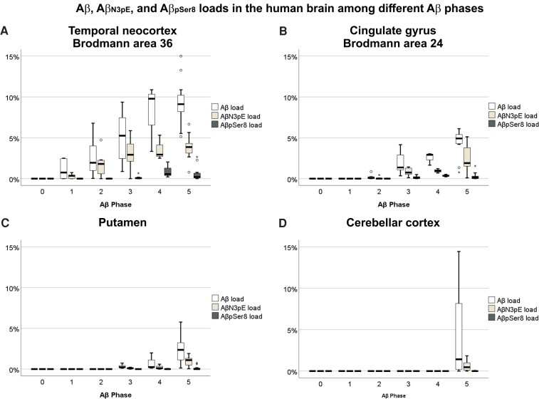
Figure 1.
Aβ, AβN3pE and AβpSer8 loads in the human brain among different Aβ phases. (A–D) Box plot diagrams comparing Aβ loads in the human brain among Aβ phases 0–5. Aβ, AβN3pE and AβpSer8 loads were obtained immunohistochemically in the temporal neocortex (Brodmann area 36) (A), the cingulate gyrus (Brodmann area 24) (B), the putamen (C) and the cerebellar cortex (D) (n = 44). All Aβ, AβN3pE and AβpSer8 loads increased with advancing Aβ phase as dependent variable (linear regression analysis with age and sex as additional covariates: P < 0.05; β: 0.289–0.731), except for the cerebellar AβpSer8 load (linear regression analysis with age and sex as additional covariates: P = 0.233). Presumably non-modified Aβ prevailed over AβN3pE and AβpSer8 in the temporal cortex, cingulate gyrus and putamen [Friedman test corrected for multiple testing (two-sided): P < 0.05]. AβN3pE was here more abundant than AβpSer8 [Friedman test corrected for multiple testing (two-sided): P < 0.05]. In the cerebellar cortex, which was only involved in Aβ pathology in 10 out of 11 Aβ phase 5 cases, presumably non-modified Aβ was more abundant than AβpSer8 [Friedman test corrected for multiple testing (two-sided): P < 0.001] and a trend was observed for more abundant AβN3pE than AβpSer8 [Friedman test corrected for multiple testing (two-sided): P = 0.059; non-corrected P = 0.019]. Box elements: centre line = median; box limits = upper and lower quartiles; whiskers = 1.5× interquartile range; dots/stars = outliers.

