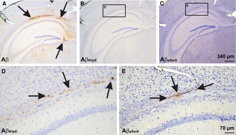
Figure 4.
Seeded Aβ in mice receiving the dispersible fraction from p-preAD case 14. (A) Seeded plaques stained with a non-C terminus-specific polyclonal antibody raised against Aβ1−40. The Aβ deposits were easily detectable even at low magnification level. (B and C) At low magnification level AβN3pE- and AβpSer8-positive plaques were less widespread and less visible. Calibration bar in C is valid for A–C. (D and E) At high power magnification both AβN3pE and AβpSer8 was seen at the white matter next to the hippocampal sector CA1. Note that AβN3pE-positive material was more widely distributed compared to AβpSer8. Calibration bar in E is valid for D and E.

