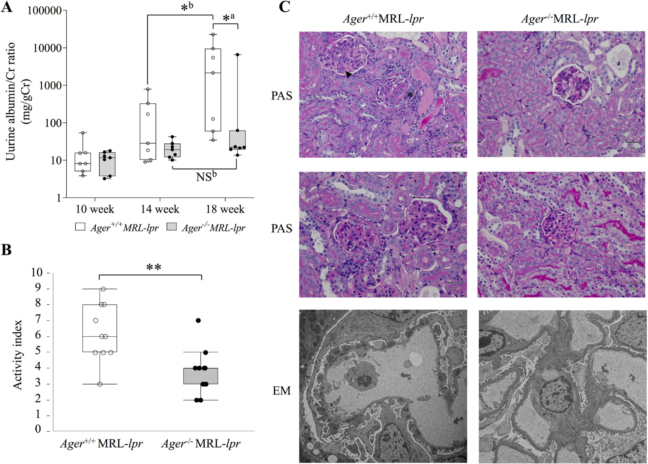
Figure 2. Attenuation of nephritis in Ager−/− MRL-lpr mice at 18 weeks of age.

(A) The transition of the urine albumin/Cr ratio (mg/gCr). Ager+/+ MRL-lpr (n = 7), and Ager−/− MRL-lpr (n = 7). aThe Mann‒Whitney U test for between-group comparisons; bWilcoxon signed-rank sum test for comparisons between 14 and 18 weeks. *P < 0.05. (B) Activity index. Ager+/+ MRL-lpr (n = 10), and Ager−/− MRL-lpr (n = 11). Student’s t-test. **P < 0.01. (C) Pathological findings of the kidney. Ager+/+ MRL-lpr shows cellular crescent (*) and karyorrhexis (arrowhead) of glomeruli (top: PAS staining). Moderate glomerular cell proliferation was found in Ager+/+ MRL-lpr mice (middle: PAS staining). By electron microscopy, subepithelial and subendothelial electron-dense deposits and podocyte foot process effacement were observed in Ager+/+ MRL-lpr mice (bottom). Representative images of Ager+/+ MRL-lpr (n = 10), and Ager−/− MRL-lpr (n = 11). NS, not significant, EM: electron microscopy; PAS, periodic acid-Schiff.
