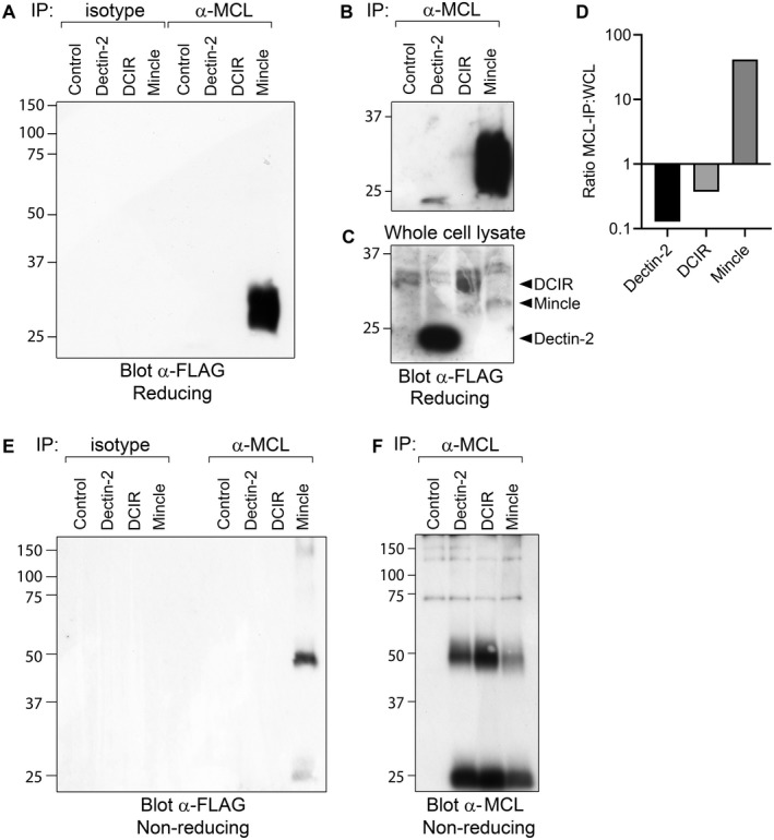
FIGURE 4.

Co‐immunoprecipitation of Mincle with MCL. 293T cells transfected with MCL and optimal amounts of FcεRIγ, together with FLAG‐Dectin‐2, FLAG‐DCIR or FLAG‐Mincle were lysed, immunoprecipitated with anti‐MCL or isotype control antibody, and separated on 10% SDS‐PAGE gels. A, Western blot analysis of immunoprecipitates under reducing conditions with anti‐FLAG antibody. A longer exposure time of the same blot is shown B. C, Whole cell lysates blotted with anti‐FLAG antibody. ImageJ was used to compare band intensities, and the ratio of specific band density in immunoprecipitation versus whole cell lysate is shown D. Immunoprecipitates were also separated under non‐reducing conditions, blotted, and detected with anti‐FLAG E or anti‐MCL F, antibodies
