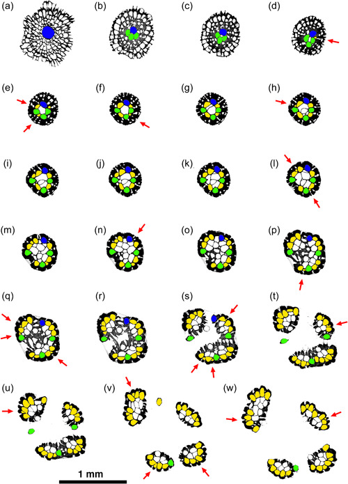
Figure 4.

Sequence of selected micro‐computed tomography transverse orthoslices upwards through the ancestrula, basal stem, and crown of a small colony of Hornera sp. Colors: blue, ancestrular zooid; green, periancestrular autozooids; yellow, exomurally budded lateral autozooids; white, frontal autozooids, kenozooids budded from the endozone and/or secondary kenozooids (outer layer). Each red arrow indicates the addition of a new exomurally budded lateral autozooid in the sequence. Sequence orthoslices (a–w) are discussed in main text
