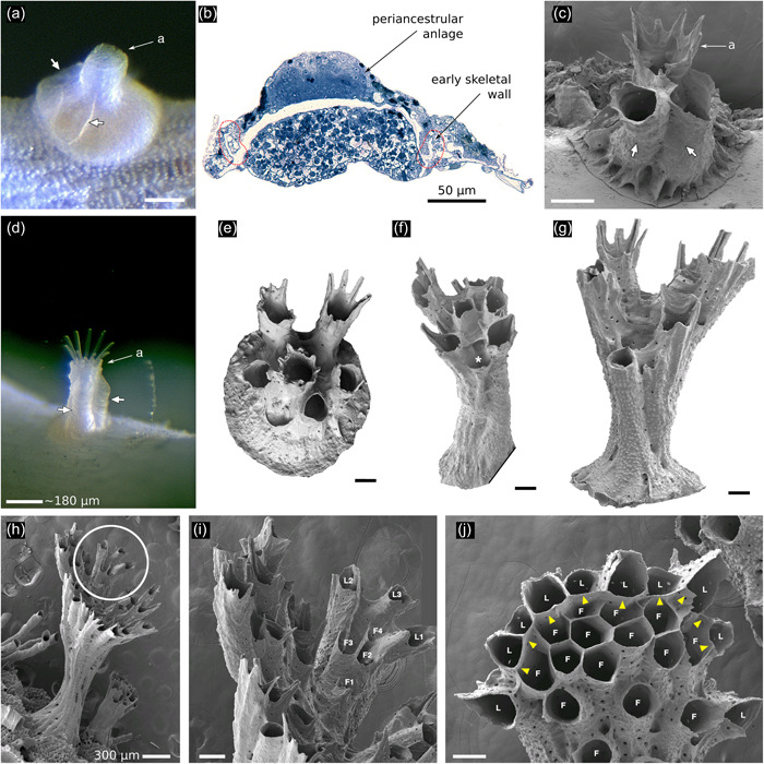
Figure 5.

Astogeny in Hornera spp. (a) Living ancestrula of Hornera sp., settled in the laboratory. White arrows show first‐formed walls of two frontally budded adventitious (periancestrular) autozooids on the ancestrula. Proximal footprint of each periancestrular zooid is approximately the same as a typical autozooid (a, aperture of ancestrula). (b) Semithin section of the same ancestrula shown in (a) with the anlage of the periancestrular zooid atop the newly calcified ancestrular dome (longitudinal section, position roughly corresponding to short arrows in a). (c) Scanning electron microscopy of more‐advanced ancestrula. White arrows show two periancestrular zooids partly fused with ancestrular tube (a). (d) Live colony showing two periancestrular zooids (white arrows) growing up wall of central ancestrular zooid (a). (e) Ancestrula of Hornera sp. 2 from Foveaux Strait. Daughter autozooids are fully separated from each other. (f) Multizooidal stem, with new exomurally budded lateral autozooid (asterisk). (g) Incipient branch crown; diverging zooids are connected by septa, long zooids with peristomial spines are laterals; central space where frontal autozooids will bud centripetally is appearing. (h) Young branch crown of Hornera sp. (i) Close up of circled region in (h), showing first‐formed lateral autozooids (L1–L3); the frontal zooids (F1–F4) bud upon and grow along the frontal budding lamina formed by the basal surface of the laterals. (j) Tip of mature branch of Hornera sp. Paramedial budding lamina is established (yellow arrowheads), frontals (F) and laterals (L) labeled. Note roofs of lateral autozooids alternate in size and distance from branch tip. Unlabeled scale bars, 100 µm
