Figure 8.

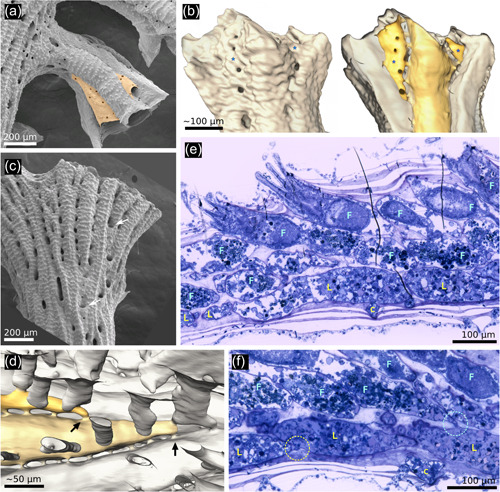
Exomural budding in Hornera. (a) Conspicuous exomural budding of two autozooids (pale orange) on a secondarily formed (=lateral) branch of Hornera robusta (abfrontal view). (b) Exterior (left) and interior (right) micro‐computed tomography reconstructions of the same Hornera sp. 1 branch tip showing two newly budded lateral autozooids (*). Viewed externally, the location of zooids is obscured by secondary calcification. (c) Abfrontal view of H. robusta branch tip, arrows show newly budded kenozooids in sulci, similar to, but more proximally sited, than exomurally budded autozooids. (d) Hypostegal pore‐associated exomural budding sites of two lateral autozooids of Hornera sp. 1; distal at left; some cancellus openings outlined for clarity. (e) Sagittal semithin section of distal branch of Hornera sp. 2 showing bilaminate zooid arrangement. Lateral (L) and frontal (F) autozooids comprise two layers of separately budded chambers interfacing at paramedial budding lamina. (f) Sagittal semithin section of H. sp. 2. Circle at left indicates hypostegal pore‐associated exomural budding site of lateral autozooid; circle at right shows an interzooidal pore‐associated budding site of frontal autozooid
