
Figure 14.
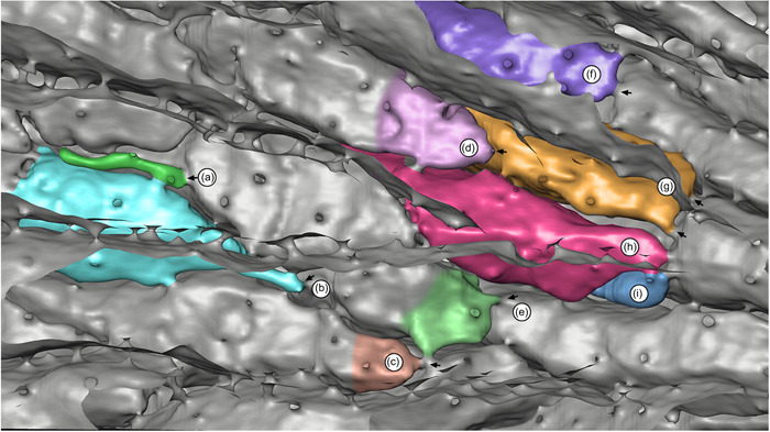
Interior reconstruction of Hornera robusta: view through the paramedial budding lamina showing the origins of frontal autozooidal chambers (partly or fully colorized for clarity); distal at left. Arrows indicate interzooidal pores at chamber origins: (a) probable aborted autozooid; (b–i) frontal autozooids. Note variability in autozooid morphology, including zooids (g–i), which originate in parallel and share proximal connections to a single frontal autozooid. Image: back‐face isosurface render through abfrontally truncated micro‐computed tomography data set