
Fig. 2. Increase in Aspn expression in response to cardiac ischemia-reperfusion injury.
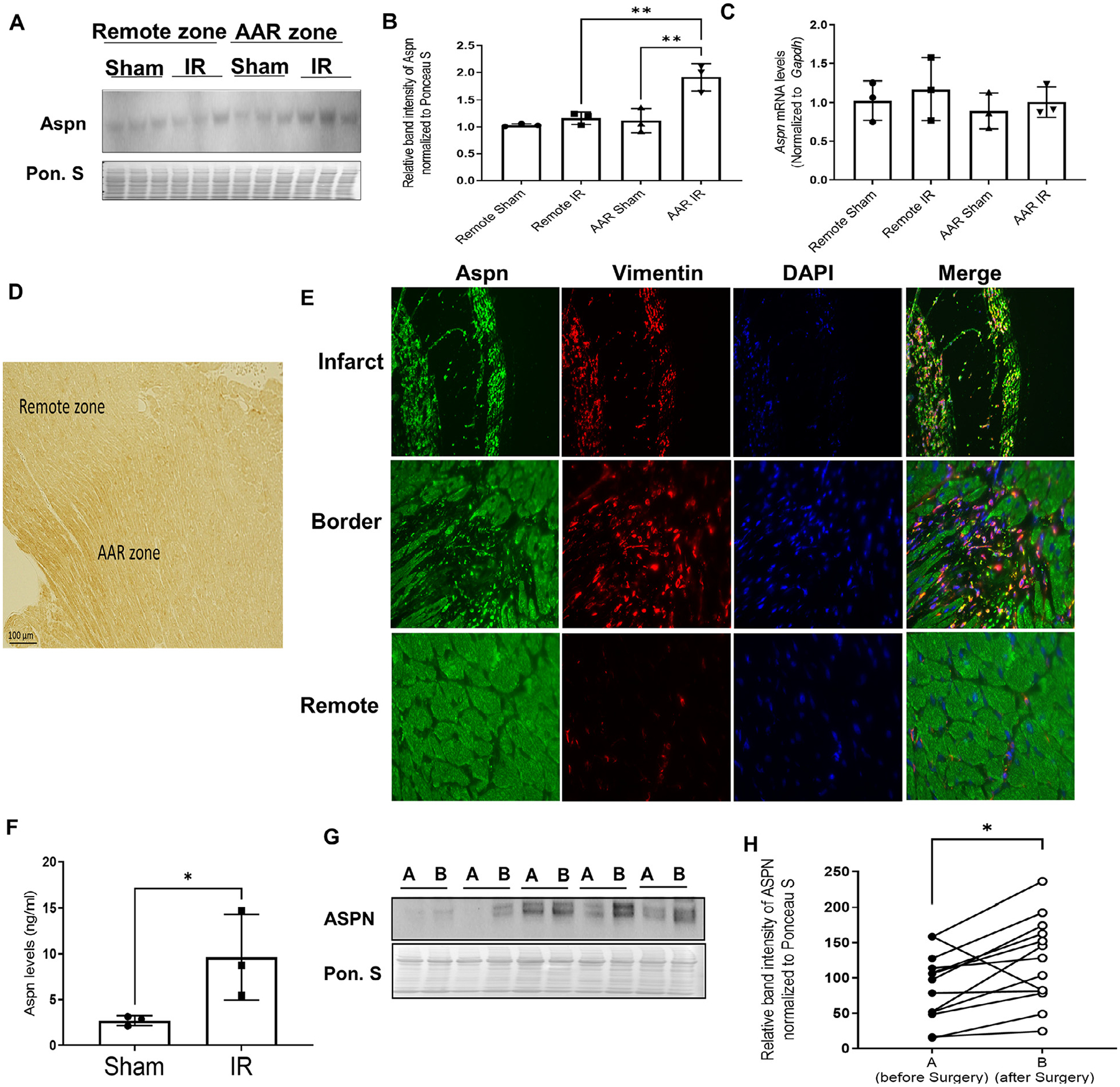
(A) Wild type C57 mice subjected to ischemia (30 mins) – reperfusion (24 hrs) injury and mice were sacrificed to separate remote zone and area at risk (AAR) zone from mice hearts. Western blot of tissue lysates for Aspn. Ponceau S staining was used to normalize the protein expression; (B) Bar graphs representing the expression of Aspn normalized to Ponceau S (n = 3); (C) qPCR analysis done on remote and AAR zone for Aspn expression normalized to GAPDH (n = 3). One-way ANOVA with Tukey multiple comparisons test was employed, **p<0.01; (D) Representative image for IHC staining on the mice heart subjected to ischemia-reperfusion injury; (E) Representative images of infarct, border and remote zone from mice heart subjected to ischemia-reperfusion injury model, co-stained with Aspn (Green), Vimentin (Red) and DAPI (blue). (F) Bar graph showing the Aspn levels from serum of the mice as detected by ELISA (n = 3). Data are expressed as mean with SD, *p ≤ 0.05 by unpaired t-test; (G) Representative western blot for Aspn and corresponding ponceau stain from tissue lysates of human atrial heart biopsies obtained before and after cardiac surgery involving cardiopulmonary bypass (CPB) and cardioplegia; (H) Quantitation of A before (A, solid circles) and after (B, empty circles) CPB (n = 13); *p<0.05 by paired t-test.