
Fig. 6. A humanized version of anti–CTLA-4×SIRPα heterodimer depletes Treg cells in a humanized mouse tumor model.
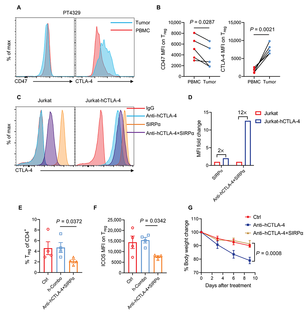
(A and B) Representative flow cytometry histograms (A) and quantification pooled from different patients (B) show CD47 and CTLA-4 expression on Treg cells of PBMCs and tumor tissues from patients with NSCLC. (C and D) Representative flow cytometry histogram (C) shows anti–hCTLA-4, SIRPα, and anti–hCTLA-4×SIRPα binding on Jurkat and Jurkat-hCTLA-4–expressing cells. Anti–hCTLA-4×SIRPα binding on Jurkat and Jurkat-hCTLA-4 cells was quantified on the basis of MFI (D). (E and F) PBMC-humanized NSG mice were inoculated with 2.5 × 10⁶ A549 tumor cells and treated with anti–hCTLA-4 plus SIRPα (h-Combo, 18 + 12 μg) or anti–hCTLA-4×SIRPα (30 μg) on day 12. Ctrl mice were treated with hIgG. Two days later, the frequency of tumor-infiltrating Treg cells of total CD4 T cells (E) and ICOS expression on Treg cells (F) were analyzed (n = 4 mice per group). (G) PBMC-humanized NSG mice were treated with 200 μg of anti–hCTLA-4 or anti–hCTLA-4×SIRPα twice a week for four times. Ctrl mice were treated with hIgG. Mouse body weight was monitored every 3 days to measure evidence of systemic toxicity (n = 5 mice per group). Data are shown as means ± SEM from two independent experiments. P values were determined by paired t test (B), Kruskal-Wallis test with Dunn’s comparison test (E), one-way ANOVA with Tukey’s multiple comparisons test (F), or two-way ANOVA with Geisser-Greenhouse’s correction (G). The normality of data was evaluated by Shapiro-Wilk test.