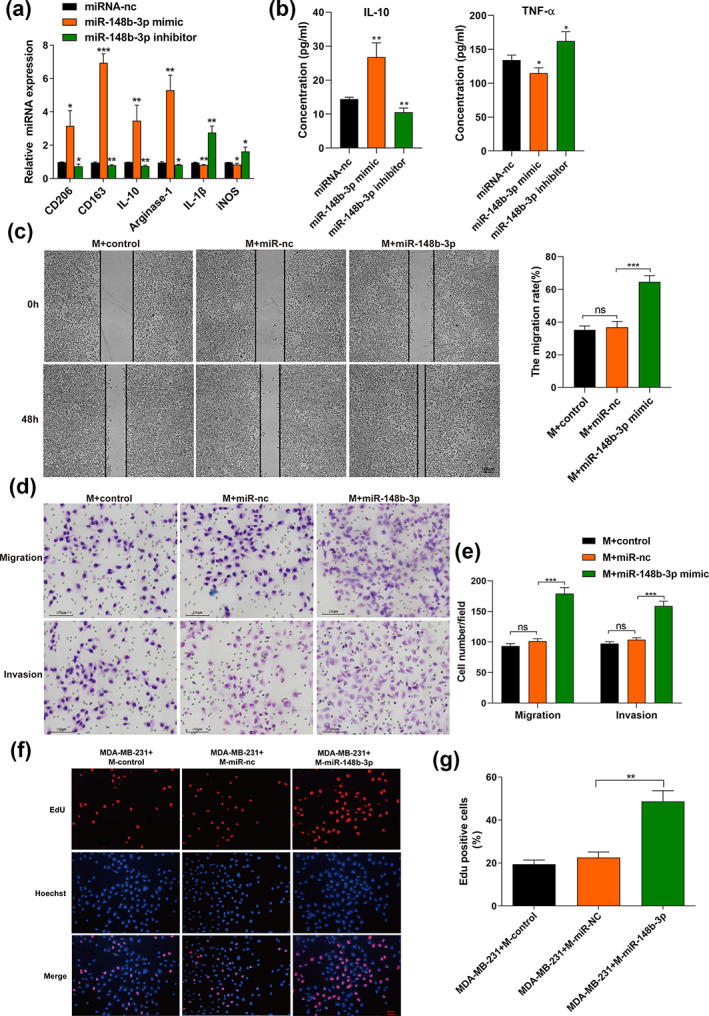
FIGURE 4.

The overexpression of exosomal miR‐148b‐3p modulates macrophage polarization and promotes proliferation, migration and invasion of breast cancer cells. (a) Macrophages were transfected with miRNA‐nc, miR‐148b‐3p mimic or miR‐148b‐3p inhibitor. After 48 h, qRT‐PCR was applied to detect the expression of M2 markers (CD163, CD206, IL‐10, arginase‐1) and M1 markers (IL‐1β, iNOS). (b) The identical method used in (a) was applied to macrophages. The different conditioned macrophage supernatants were harvested to detect the release of IL‐10 and TNF‐α by ELISA. (c) Scratch experiment. Notably, we used mitomycin C (10 μg/mL) to treat MDA‐MB‐231 cells for 2 h before conducting the scratch assay. After scratching, the cells were washed with PBS and replaced with conditioned medium from macrophages transfected with miR‐nc, miR‐148b‐3p mimic or not. Images were taken at 0 and 48 h (scale bar, 100 μm). Original magnification ×100. “M+ miR‐148b‐3p” refers to “macrophages treated with miR‐148b‐3p mimic”. (d, e) Transwell assay was performed to evaluate the migration and invasion of MDA‐MB‐231 cells. Macrophages transfected with miR‐nc, miR‐148b‐3p mimic and then cultured with MDA‐MB‐231 cells in the indirect coculture system in vitro. The control group was not transfected with any exogenous miRNA. After incubation for 24 h, MDA‐MB‐231 cells were detached from the upper chamber, and the migration and invasion ability of MDA‐MB‐231 cells was detected. Illustrative images of migratory or invasive cells and quantifications are shown. Scale bar, 100 μm. Original magnification ×100. (f, g) EdU assay was performed to evaluate the proliferation of MDA‐MB‐231 cells. MDA‐MB‐231 cells were cocultured with conditioned macrophages as described in (a) for 48 h, and the positive cell ratio is shown. Scale bar, 50 μm. Original magnification ×200. The student's t‐test was adopted to analyze the statistical significance of the difference between two groups, and one‐way ANOVA test was utilized to analyze multiple groups (*p < 0.05, **p < 0.01, ***p < 0.001).
