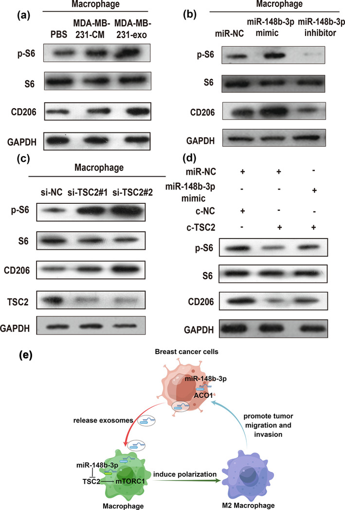
FIGURE 8.

Exosomal miR‐148b‐3p regulates macrophage polarization by inhibiting TSC2 and activating the mTORC1 signaling pathway. (a) Western blot (WB) analysis of mTORC1 activity markers (p‐S6, S6) and M2‐related gene CD206 expression in M0 macrophages treated with PBS, MDA‐MB‐231‐CM, MDA‐MB‐231‐exo or (b) miR‐NC, miR‐148b‐3p mimic, miR‐148b‐3p inhibitor. (c) WB assay of mTORC1 activity markers (p‐S6, S6), TSC2 and M2‐related gene CD206 expression in M0 macrophages treated with si‐NC as control, si‐TSC2#1 and si‐TSC2#2 for downregulating TSC2 expression. (d) WB analysis of mTORC1 activity markers (p‐S6, S6) and M2‐related gene CD206 expression in M0 macrophages treated with the indicated treatment. Each experiment was conducted at least three times. (e) Schematic diagram of exosomal miR‐148b‐3p promoted M2 macrophage polarization via the TSC2/mTORC1 signaling pathway was drawn using Figdraw.
