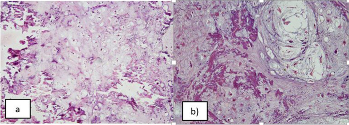
Fig. 6.
a Haematoxylin and eosin stained section, 100x: shows a tumour composed of lobules of chondrocytic cells within lacunae; left half of the image shows presence of osteoid matrix. b Haematoxylin and eosin stained section, 200X: Higher magnification shows chondroid areas containing tumour cells in lacunar spaces that are intimately associated with osteoid matrix (darker pink) present in the left half of the image

